 Explore
 Validate
 Learn
Learn Western blot
 Immunohistochemistry
Antibody data
- Antibody Data
- Antigen structure
- References [0]
- Comments [0]
- Validations
- Western blot [2]
- Immunohistochemistry [7]
Submit
Validation data
Reference
Comment
Report error
- Product number
- AMAb91418 - Provider product page

- Provider
- Atlas Antibodies
- Proper citation
- Atlas Antibodies Cat#AMAb91418, RRID:AB_2732085
- Product name
- Anti-BCL11B
- Antibody type
- Monoclonal
- Antigen
- Synthetic peptide
- Reactivity
- Human, Mouse
- Host
- Mouse
- Conjugate
- Unconjugated
- Antigen sequence
EIGIQVTPDEDDHLLSPTK- Isotype
- IgG
- Antibody clone number
- CL6426
- Vial size
- 100 µl
- Storage
- Store at +4°C for short term storage. Long time storage is recommended at -20°C.
No comments: Submit comment
Supportive validation
- Submitted by
- Atlas Antibodies (provider)
- Main image

- Experimental details
- Western blot analysis in human cerebral cortex tissue.

- Submitted by
- Atlas Antibodies (provider)
- Main image

- Experimental details
- Western blot analysis in mouse cerebral cortex tissue.

Enhanced validation
Supportive validation
- Submitted by
- Atlas Antibodies (provider)
- Enhanced method
- Orthogonal validation
- Main image
- Experimental details
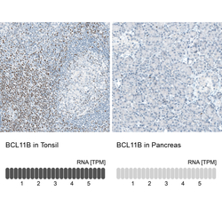
- Immunohistochemistry analysis in human tonsil and pancreas tissues using AMAb91418 antibody. Corresponding BCL11B RNA-seq data are presented for the same tissues.
- Sample type
- HUMAN

Supportive validation
- Submitted by
- Atlas Antibodies (provider)
- Main image
- Experimental details
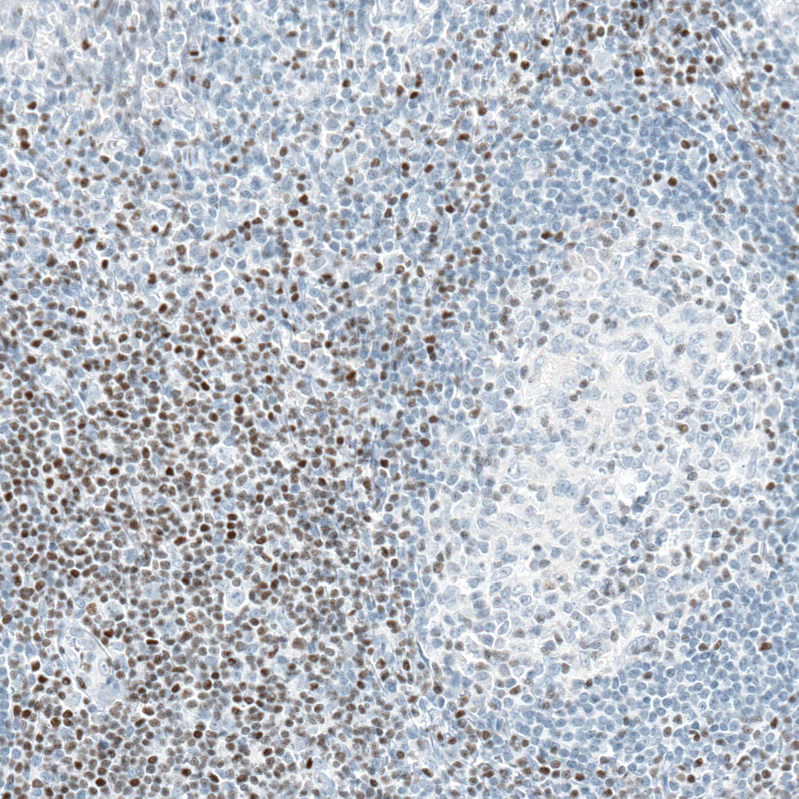
- Immunohistochemical staining of human tonsil shows moderate to strong nuclear positivity, mainly in non - germinal center cells.
- Submitted by
- Atlas Antibodies (provider)
- Main image
- Experimental details
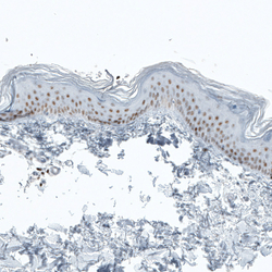
- Immunohistochemical staining of human skin shows moderate to strong nuclear positivity in dermal cells.

- Submitted by
- Atlas Antibodies (provider)
- Main image

- Experimental details
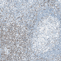
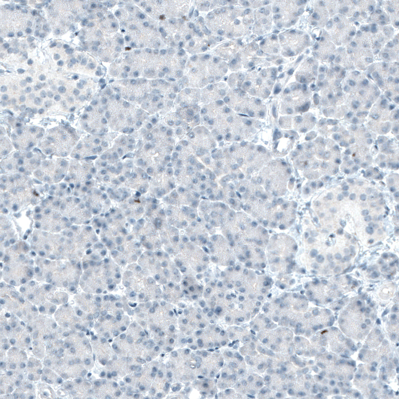
- Immunohistochemical staining of human pancreas shows no positivity in exocrine glandular cells as expected.
- Submitted by
- Atlas Antibodies (provider)
- Main image
- Experimental details
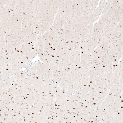
- Immunohistochemical staining of mouse cerebral cortex shows moderate to strong nuclear positivity in neurons, mainly in layers 5-6.

- Submitted by
- Atlas Antibodies (provider)
- Main image
- Experimental details
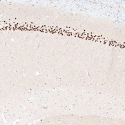
- Immunohistochemical staining of mouse hippocampus shows strong nuclear positivity in neurons in CA1 pyramidal layer.

- Submitted by
- Atlas Antibodies (provider)
- Main image
- Experimental details
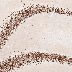
- Immunohistochemical staining of mouse hippocampus shows strong nuclear positivity in neurons in the granular cell layer of dentate gyrus.
