Explore
 Validate
 Learn
Learn Western blot
Antibody data
- Antibody Data
- Antigen structure
- References [1]
- Comments [0]
- Validations
- Western blot [1]
- Immunocytochemistry [1]
- Immunohistochemistry [6]
- Other assay [1]
Submit
Validation data
Reference
Comment
Report error
- Product number
- PA5-57077 - Provider product page

- Provider
- Invitrogen Antibodies
- Product name
- SF3B14 Polyclonal Antibody
- Antibody type
- Polyclonal
- Antigen
- Recombinant full-length protein
- Description
- Immunogen sequence: YEDIFDAKNA CDHLSGFNVC NRYLVVLYYN ANRAFQKMDT KKKEEQLKLL KEKYGINTDP P
- Concentration
- 0.10 mg/mL
Submitted references The long non-coding RNA DKFZp434J0226 regulates the alternative splicing process through phosphorylation of SF3B6 in PDAC.
Li J, Tong H, Li D, Jiang Q, Zhang Y, Tang W, Jin D, Chen S, Qin X, Zhang S, Xue R
Molecular medicine (Cambridge, Mass.) 2021 Aug 28;27(1):95
Molecular medicine (Cambridge, Mass.) 2021 Aug 28;27(1):95
No comments: Submit comment
Supportive validation
- Submitted by
- Invitrogen Antibodies (provider)
- Main image

- Experimental details
- Western blot analysis of SF3B6 in Lane 1: NIH-3T3 cell lysate (Mouse embryonic fibroblast cells); Lane 2: NBT-II cell lysate (Rat Wistar bladder tumour cells). Samples were probed using a SF3B6 Polyclonal Antibody (Product # PA5-57077).

Supportive validation
- Submitted by
- Invitrogen Antibodies (provider)
- Main image

- Experimental details
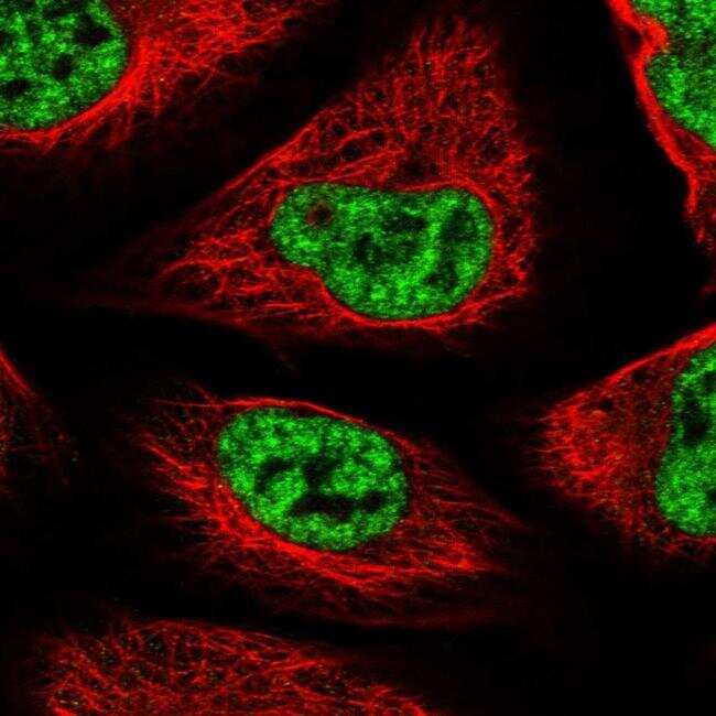
- Immunofluorescent staining of SF3B6 in human cell line U-2 OS shows positivity in nucleus but excluded from the nucleoli. Samples were probed using a SF3B6 Polyclonal Antibody (Product # PA5-57077).
Supportive validation
- Submitted by
- Invitrogen Antibodies (provider)
- Main image

- Experimental details
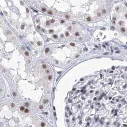
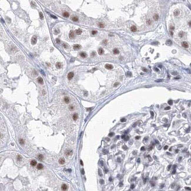
- Immunohistochemical staining of SF3B14 in human lymph node using SF3B14 Polyclonal Antibody (Product # PA5-57077).

- Submitted by
- Invitrogen Antibodies (provider)
- Main image
- Experimental details
- Immunohistochemical staining of SF3B14 in human kidney using SF3B14 Polyclonal Antibody (Product # PA5-57077).
- Submitted by
- Invitrogen Antibodies (provider)
- Main image

- Experimental details
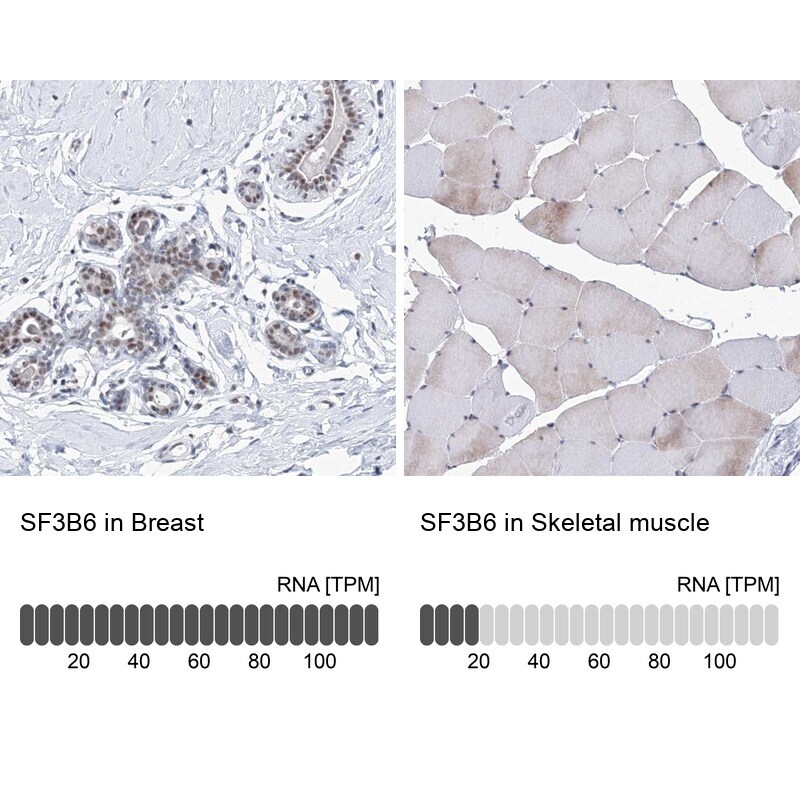
- Immunohistochemical staining of SF3B14 in human breast and skeletal muscle tissues using SF3B14 Polyclonal Antibody (Product # PA5-57077). Corresponding SF3B6 RNA-seq data are presented for the same tissues.
- Submitted by
- Invitrogen Antibodies (provider)
- Main image
- Experimental details
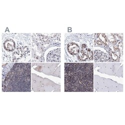
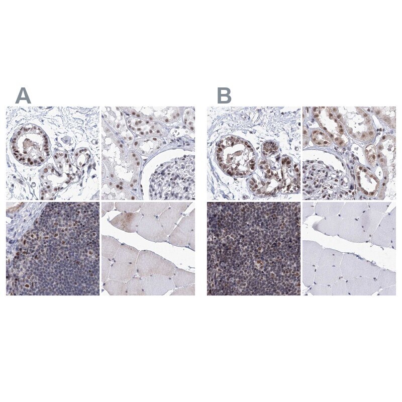
- Immunohistochemical staining of SF3B14 in human breast, kidney, lymph node and skeletal muscle using SF3B14 Polyclonal Antibody (Product # PA5-57077) (A) shows similar protein distribution across tissues to an independent SF3B14 Polyclonal Antibody (B).
- Submitted by
- Invitrogen Antibodies (provider)
- Main image

- Experimental details
- Immunohistochemical staining of SF3B14 in human skeletal muscle using SF3B14 Polyclonal Antibody (Product # PA5-57077) shows low expression as expected.

- Submitted by
- Invitrogen Antibodies (provider)
- Main image

- Experimental details
- Immunohistochemical staining of SF3B14 in human breast using SF3B14 Polyclonal Antibody (Product # PA5-57077) shows high expression.

Supportive validation
- Submitted by
- Invitrogen Antibodies (provider)
- Main image
- Experimental details
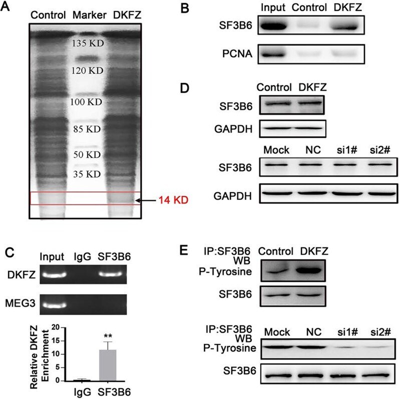
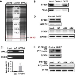
- Fig. 5 SF3B6 serves as a DKFZp434J0226 (DKFZ)-binding protein. A RNA pull-down assay performed in the HEK293T cell lysates with biotin-labeled oligos. After pull-down, proteins were subjected to SDS-PAGE and stained by Coomassie brilliant blue. The band indicated by the arrow was subjected to mass spectrometry. B Western blotting analysis determined the specific interaction of sense DKFZ with SF3B6 protein, but not with PCNA protein (negative control). C RNA immunoprecipitation (RIP) of SF3B6 interaction with DKFZ in the HEK293T cells. RNA-protein complexes immunoprecipitated by anti-SF3B6 or control IgG were determined by RT-qPCR using specific primers for DKFZ or MEG3 (negative control). Data are shown as mean +- SD (n = 4); ** P < 0.01. D Western blotting analysis of SF3B6 levels in the indicated DKFZ overexpression AsPC-1 cells or DKFZ knockdown MIAPaCa-2 cells. E Western blotting analysis of SF3B6 phosphorylation levels in the indicated DKFZ overexpression AsPC-1 cells or DKFZ knockdown MIAPaCa-2 cells. For SF3B6 phosphorylation detection, cell lysates were prepared and subjected to immunoprecipitation (IP) with anti-SF3B6 antibody, followed by immunoblotting analysis with the anti-phospho-tyrosine antibody