Explore
 Validate
 Learn
Learn Western blot
Antibody data
- Antibody Data
- Antigen structure
- References [4]
- Comments [0]
- Validations
- Western blot [2]
- Immunocytochemistry [3]
Submit
Validation data
Reference
Comment
Report error
- Product number
- PA1-767A - Provider product page

- Provider
- Invitrogen Antibodies
- Product name
- VAMP3 Polyclonal Antibody
- Antibody type
- Polyclonal
- Antigen
- Synthetic peptide
- Description
- PA1-767A detects vesicle-associated membrane protein 3 (VAMP-3)/cellubrevin in human, rat and mouse samples. PA1-767A has been successfully used in Western blot and ICC/IF procedures. By Western blot, this antibody detects an ~13 kDa protein representing VAMP-3 in HeLa cell lysates. PA1-767A immunogen is a sythetic peptide corresponding to residues M(1) S T G V P S G S S A A T G S N R R(18) C of mouse VAMP-3. This immunizing peptide (Cat. # PEP-145) is available for use in neutralization and control experiments.
- Reactivity
- Human, Mouse, Rat
- Host
- Rabbit
- Isotype
- IgG
- Vial size
- 100 µg
- Concentration
- 1 mg/mL
- Storage
- -20° C, Avoid Freeze/Thaw Cycles
Submitted references Single bioengineered ncRNA molecule for dual-targeting toward the control of non-small cell lung cancer patient-derived xenograft tumor growth.
Recycling endosomes attach to the trans-side of Golgi stacks in Drosophila and mammalian cells.
SNAP23, Syntaxin4, and vesicle-associated membrane protein 7 (VAMP7) mediate trafficking of membrane type 1-matrix metalloproteinase (MT1-MMP) during invadopodium formation and tumor cell invasion.
Characterization of VAMP-2 in the lung: implication in lung surfactant secretion.
Petrek H, Yan Ho P, Batra N, Tu MJ, Zhang Q, Qiu JX, Yu AM
Biochemical pharmacology 2021 Jul;189:114392
Biochemical pharmacology 2021 Jul;189:114392
Recycling endosomes attach to the trans-side of Golgi stacks in Drosophila and mammalian cells.
Fujii S, Kurokawa K, Inaba R, Hiramatsu N, Tago T, Nakamura Y, Nakano A, Satoh T, Satoh AK
Journal of cell science 2020 Feb 26;133(4)
Journal of cell science 2020 Feb 26;133(4)
SNAP23, Syntaxin4, and vesicle-associated membrane protein 7 (VAMP7) mediate trafficking of membrane type 1-matrix metalloproteinase (MT1-MMP) during invadopodium formation and tumor cell invasion.
Williams KC, McNeilly RE, Coppolino MG
Molecular biology of the cell 2014 Jul 1;25(13):2061-70
Molecular biology of the cell 2014 Jul 1;25(13):2061-70
Characterization of VAMP-2 in the lung: implication in lung surfactant secretion.
Wang P, Howard MD, Zhang H, Chintagari NR, Bell A, Jin N, Mishra A, Liu L
Cell biology international 2012 Sep;36(9):785-91
Cell biology international 2012 Sep;36(9):785-91
No comments: Submit comment
Supportive validation
- Submitted by
- Invitrogen Antibodies (provider)
- Main image
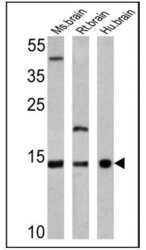
- Experimental details
- Western blot analysis of VAMP3 was performed by loading 25 µg of mouse brain (lane 1), rat brain (lane 2) and human brain (lane 3) lysates onto an SDS polyacrylamide gel. Proteins were transferred to a PVDF membrane and blocked at 4ºC overnight. The membrane was probed with a VAMP3 polyclonal antibody (Product # PA1-767A) at a dilution of 1:2000 overnight at 4°C, washed in TBST, and probed with an HRP-conjugated secondary antibody for 1 hr at room temperature in the dark. Chemiluminescent detection was performed using Pierce ECL Plus Western Blotting Substrate (Product # 32132). Results show a band at ~13 kDa.

- Submitted by
- Invitrogen Antibodies (provider)
- Main image
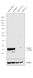
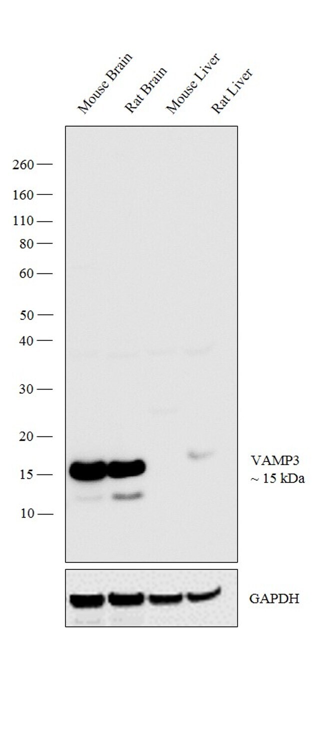
- Experimental details
- Western blot analysis was performed on tissue extracts (30 µg lysate) of Mouse Brain (Lane 1), Rat Brain (Lane 2), Mouse Liver (Lane 3) and Rat Liver (Lane 4). The blot was probed with Anti-VAMP3 Polyclonal Antibody (Product # PA1-767A, 1:2000 dilution) and detected by chemiluminescence using Goat anti-Rabbit IgG (H+L) Superclonal™ Secondary Antibody, HRP conjugate (Product # A27036, 0.25 µg/ml, 1:4000 dilution). A 15 kDa band corresponding to VAMP3 was observed across the tissue extracts tested except Mouse Liver and Rat Liver tested which is reported to be negative.
Supportive validation
- Submitted by
- Invitrogen Antibodies (provider)
- Main image

- Experimental details
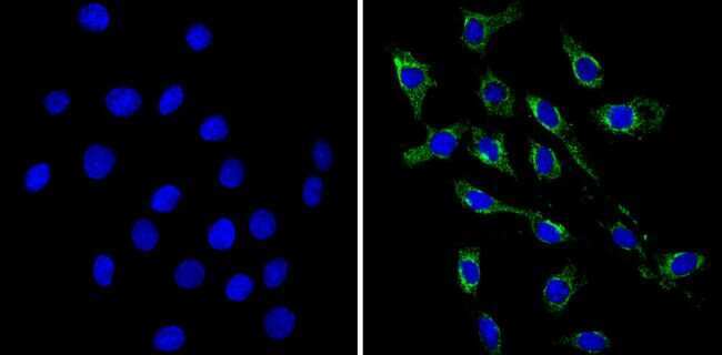
- Immunofluorescent analysis of VAMP3 (green) showing staining in the cytoplasm of C6 cells (right) compared to a negative control without primary antibody (left). Formalin-fixed cells were permeabilized with 0.1% Triton X-100 in TBS for 5-10 minutes and blocked with 3% BSA-PBS for 30 minutes at room temperature. Cells were probed with a VAMP3 polyclonal antibody (Product # PA1-767A) in 3% BSA-PBS at a dilution of 1:50 and incubated overnight at 4ºC in a humidified chamber. Cells were washed with PBST and incubated with a DyLight-conjugated secondary antibody in PBS at room temperature in the dark. Nuclei were stained with Hoechst or DAPI (blue). Images were taken at a magnification of 60x.
- Submitted by
- Invitrogen Antibodies (provider)
- Main image

- Experimental details
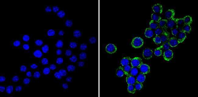
- Immunofluorescent analysis of VAMP3 (green) showing staining in the cytoplasm of Neuro-2a cells (right) compared to a negative control without primary antibody (left). Formalin-fixed cells were permeabilized with 0.1% Triton X-100 in TBS for 5-10 minutes and blocked with 3% BSA-PBS for 30 minutes at room temperature. Cells were probed with a VAMP3 polyclonal antibody (Product # PA1-767A) in 3% BSA-PBS at a dilution of 1:50 and incubated overnight at 4ºC in a humidified chamber. Cells were washed with PBST and incubated with a DyLight-conjugated secondary antibody in PBS at room temperature in the dark. Nuclei were stained with Hoechst or DAPI (blue). Images were taken at a magnification of 60x.
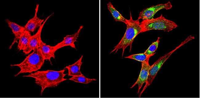
- Submitted by
- Invitrogen Antibodies (provider)
- Main image
- Experimental details
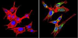
- Immunofluorescent analysis of VAMP3 (green) showing staining in the cytoplasm of NIH-3T3 cells (right) compared to a negative control without primary antibody (left). Formalin-fixed cells were permeabilized with 0.1% Triton X-100 in TBS for 5-10 minutes and blocked with 3% BSA-PBS for 30 minutes at room temperature. Cells were probed with a VAMP3 polyclonal antibody (Product # PA1-767A) in 3% BSA-PBS at a dilution of 1:50 and incubated overnight at 4ºC in a humidified chamber. Cells were washed with PBST and incubated with a DyLight-conjugated secondary antibody in PBS at room temperature in the dark. Actin was stained using Alexa Fluor 554 (red) and nuclei were stained with Hoechst or DAPI (blue). Images were taken at a magnification of 60x.