Explore
 Validate
 Learn
Learn Western blot
Antibody data
- Antibody Data
- Antigen structure
- References [2]
- Comments [0]
- Validations
- Western blot [1]
- Other assay [4]
Submit
Validation data
Reference
Comment
Report error
- Product number
- PA5-42099 - Provider product page

- Provider
- Invitrogen Antibodies
- Product name
- SEMG2 Polyclonal Antibody
- Antibody type
- Polyclonal
- Antigen
- Synthetic peptide
- Description
- Peptide sequence: GQKGQHYFGQ KDQQHTKSKG SFSIQHTYHV DINDHDWTRK SQQYDLNALH
- Concentration
- 0.5 mg/mL
Submitted references Cancer-testis antigens, semenogelins 1 and 2, exhibit different anti-proliferative effects on human lung adenocarcinoma cells.
SEMG1/2 augment energy metabolism of tumor cells.
Shuvalov O, Kizenko A, Petukhov A, Aksenov N, Fedorova O, Vorobev M, Daks A, Barlev N
Cell death discovery 2020;6:108
Cell death discovery 2020;6:108
SEMG1/2 augment energy metabolism of tumor cells.
Shuvalov O, Kizenko A, Petukhov A, Fedorova O, Daks A, Bottrill A, Snezhkina AV, Kudryavtseva AV, Barlev N
Cell death & disease 2020 Dec 11;11(12):1047
Cell death & disease 2020 Dec 11;11(12):1047
No comments: Submit comment
Supportive validation
- Submitted by
- Invitrogen Antibodies (provider)
- Main image
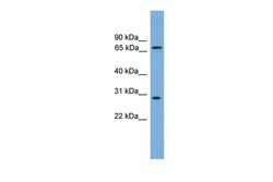
- Experimental details
- Western blot analysis of human COLO205 cell lysate using an anti-SEMG2 polyclonal antibody (Product # PA5-42099).
Supportive validation
- Submitted by
- Invitrogen Antibodies (provider)
- Main image
- Experimental details
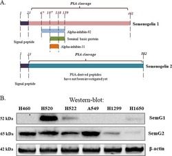
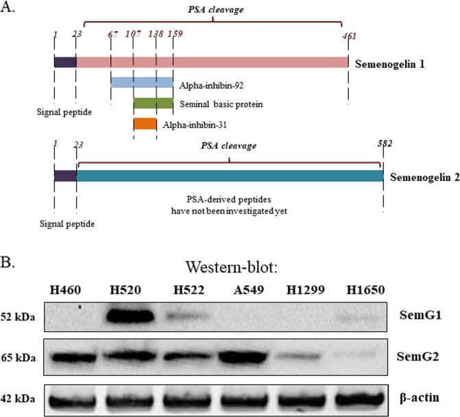
- Fig. 1 NSCLC cell lines express SEMG1 and SEMG2. a Comparison of SEMG1 and SEMG2. PSA - prostate specific antigen, cleaves SEMG1 and SEMG2 to small peptides indicated. b Distribution of SEMG1 and SEMG2 in different NSCLC cell lines. Western-blot analysis.
- Submitted by
- Invitrogen Antibodies (provider)
- Main image
- Experimental details
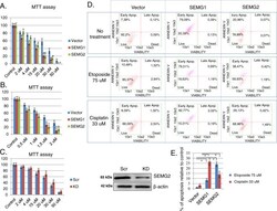
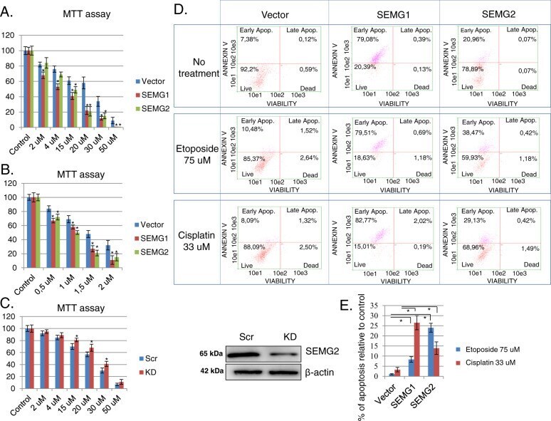
- Fig. 5 SEMGs induce apoptosis and sensitize cells to genotoxic drugs. MTT assay of H1299 cells overexpressing vector, SEMG1, SEMG2 and incubated with different concentrations of a doxorubicin or b cisplatin. c MTT assay of H1299 cells with scramble or SEMG2 knockdown (KD) treated with cisplatin. Knockdown of SEMG2 is visualized by western-blotting. d Apoptosis profile of non-treated or treated with 30 muM of cisplatin or 75 muM of etoposide H1299 cells overexpressing vector, SEMG1 or SEMG2. e Percentage of apoptosis increase in H1299 cells with vector, SEMG1 or SEMG2 overexpressing after treatment with etoposide or cisplatin. Results are represented as mean +- SEM of three experiments. ** P < 0.01.
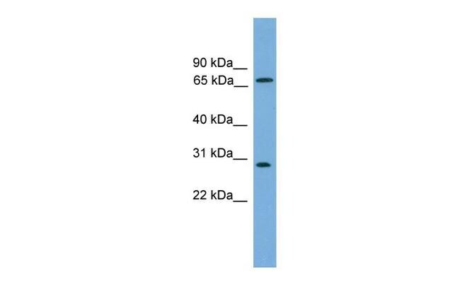
- Submitted by
- Invitrogen Antibodies (provider)
- Main image
- Experimental details
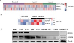
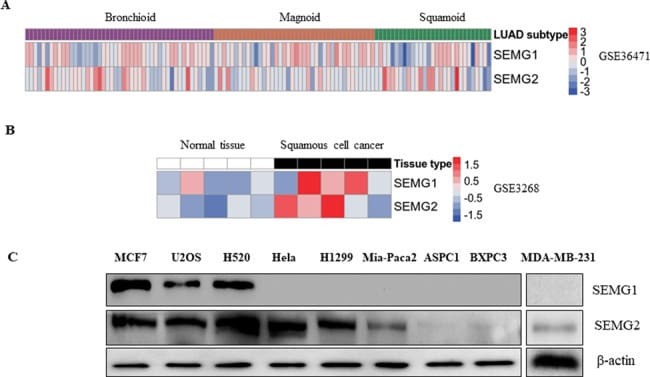
- Fig. 1 SEMG1 and SEMG2 are frequently expressed in human cancer cell lines of different origin and clinical samples. A Heat map of SEMG1 and SEMG2 expression in human lung cancer cell models (GSE36471) based on the RNA-seq data. Pre-calculated expression values (Phantasus software) were log-transformed and quantile normalized using the R (v3.61) statistical language in the R studio software (v1.2.5001). Heatmaps were produced using the ggplot2 library. B Heat map of SEMG1 and SEMG2 expression in clinical samples of lung squamous carcinoma (GSE3268) and the corresponding normal tissue. C Western-blot analysis of several human tumor cell lines of different origin for SEMG1 and SEMG2 expression.
- Submitted by
- Invitrogen Antibodies (provider)
- Main image
- Experimental details
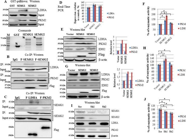
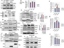
- Fig. 4 SEMG1 and SEMG2 interact with PKM2 and LDHA, upregulates their protein level and enzymatic activity. Recombinant SEMG1 and SEMG2 bind PKM2, whereas SEMG1 only binds LDHA in GST pull-down assay followed by western-blotting. B 3xFlag-tagged SEMG1 and SEMG2 bind PKM2, whereas SEMG1 only binds LDHA in co-immunoprecipitation. C 3xFlag-tagged PKM2 interacts with both endogenous SEMG1 and SEMG2 in H520 cells, whereas 3xFlag-tagged LDHA binds SEMG1 only (co-immunoprecipitation). D Overexpression of SEMG1 and SEMG2 in H1299 cell line does not alters the mRNA levels of PKM2 and LDHA (Real-Time PCR). The stable overexpression of SEMG1 and SEMG2 increase the protein level and enzymatic activity of PKM2, whereas the overexpression of SEMG1 only elevates the protein level and enzymatic activity of LDHA in E , F H1299 cells and G , H Mia-Paca 2 cells. Knockdown of SEMG1 and SEMG2 in H520 cells decreases the protein level ( I ) and enzymatic activity ( J ) of PKM2 and LDHA. Three biological replicates were used for all quantifications, data are presented as mean +- S.D., * P < 0.05.