Explore
 Validate
 Learn
Learn Western blot
Antibody data
- Antibody Data
- Antigen structure
- References [1]
- Comments [0]
- Validations
- Western blot [2]
- Immunocytochemistry [1]
- Other assay [1]
Submit
Validation data
Reference
Comment
Report error
- Product number
- PA5-22174 - Provider product page

- Provider
- Invitrogen Antibodies
- Product name
- Ubiquilin 1 Polyclonal Antibody
- Antibody type
- Polyclonal
- Antigen
- Recombinant protein fragment
- Description
- Recommended positive controls: 293T, A431, H1299, HeLa, HepG2, Molt-4, Raji.
- Concentration
- 1 mg/mL
Submitted references p62/Sequestosome 1 levels increase and phosphorylation is altered in Cx50D47A lenses, but deletion of p62/sequestosome 1 does not improve transparency.
Jara O, Mysliwiec H, Minogue PJ, Berthoud VM, Beyer EC
Molecular vision 2020;26:204-215
Molecular vision 2020;26:204-215
No comments: Submit comment
Supportive validation
- Submitted by
- Invitrogen Antibodies (provider)
- Main image

- Experimental details
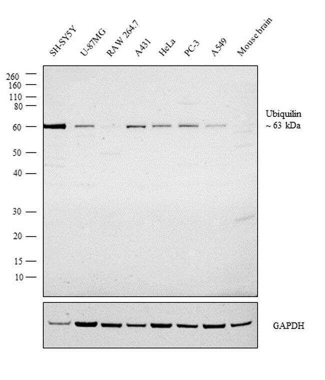
- Western blot analysis was performed on whole cell extract (30 µg lysate) of SH-SY5Y (Lane 1), U-87MG (Lane 2), RAW 264.7 (Lane 3), A431 (Lane 4), HeLa (Lane 5), PC-3 (Lane 6), A549 (Lane 7) and tissue extract of Mouse Brain (Lane 8). The blot was probed with Anti-Ubiquilin 1 Polyclonal Antibody (Product # PA5-22174, 1:3000 dilution) and detected by chemiluminescence using Goat anti-Rabbit IgG (H+L) Superclonal™ Secondary Antibody, HRP conjugate (Product # A27036, 0.25 µg/ml, 1:4000 dilution). A 63 kDa band corresponding to Ubiquilin 1 was detected in cell lines tested where as mouse origin cell line or tissue shows extremely weak signal, indicating human specificity.
- Submitted by
- Invitrogen Antibodies (provider)
- Main image

- Experimental details
- Western Blot using Ubiquilin 1 Polyclonal Antibody (Product # PA5-22174). Sample (30 µg of whole cell lysate). Lane A: 293T . 7.5% SDS PAGE. Ubiquilin 1 Polyclonal Antibody (Product # PA5-22174) diluted at 1:1,000.

Supportive validation
- Submitted by
- Invitrogen Antibodies (provider)
- Main image

- Experimental details
- Immunofluorescent analysis of Ubiquilin-1 in paraformaldehyde-fixed A549 cells using a Ubiquilin-1 polyclonal antibody (Product # PA5-22174) at a 1:200 dilution.

Supportive validation
- Submitted by
- Invitrogen Antibodies (provider)
- Main image

- Experimental details
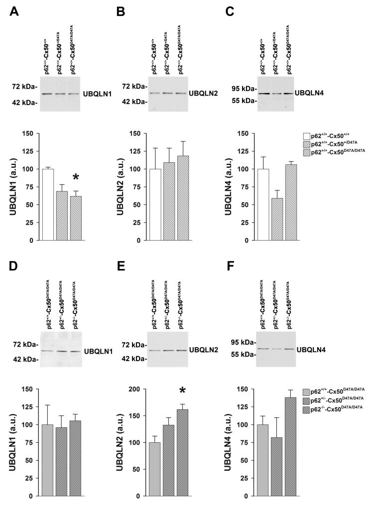
- Figure 7 Levels of ubiquilins are altered in lenses of homozygous Cx50D47A mice with or without the deletion of p62. A - C : Immunoblots show levels of UBQLN1 ( A ), UBQLN2 ( B ), and UBQLN4 ( C ) in lens homogenates prepared from 1-month-old wild-type ( p62 +/+ -Cx50 +/+ ), or heterozygous ( p62 +/+ -Cx50 +/D47A ) or homozygous ( p62 +/+ -Cx50 D47A/D47A ) Cx50D47A mice. The migration positions of the molecular mass markers are indicated on the left. The graphs show the densitometric values of the immunoreactive ubiquilin bands. Data are presented as the mean (bar) + standard error of the mean (SEM; n = 3 sets) in arbitrary units (a.u.). D - F : Immunoblots show levels of UBQLN1 ( D ), UBQLN2 ( E ), and UBQLN4 ( F ) in lens homogenates prepared from 1-month-old homozygous Cx50D47A mice that were wild-type ( p62 +/+ -Cx50 D47A/D47A ) or heterozygous ( p62 +/- -Cx50 D47A/D47A ) or homozygous ( p62 -/- -Cx50 D47A/D47A ) p62 . The migration positions of the molecular mass markers are indicated on the left. The graphs show the densitometric values of the immunoreactive ubiquilin bands. Data are presented as the mean (bar) + SEM (n = 3 sets) in arbitrary units (a.u.). Asterisks denote statistically significant differences from wild-type lenses (p