 Explore
 Validate
 Learn
Learn Western blot
Antibody data
- Antibody Data
- Antigen structure
- References [3]
- Comments [0]
- Validations
- Western blot [4]
- Immunohistochemistry [4]
Submit
Validation data
Reference
Comment
Report error
- Product number
- NBP1-82479 - Provider product page

- Provider
- Novus Biologicals
- Proper citation
- Novus Cat#NBP1-82479, RRID:AB_11010467
- Product name
- Rabbit Polyclonal Sigma-1 R/OPRS1 Antibody
- Antibody type
- Polyclonal
- Description
- Immunogen affinity purified. Specificity of human Sigma-1 R/OPRS1 antibody verified on a Protein Array containing target protein plus 383 other non-specific proteins.
- Reactivity
- Human
- Host
- Rabbit
- Isotype
- IgG
- Vial size
- 0.1 ml
- Storage
- Store at 4C short term. Aliquot and store at -20C long term. Avoid freeze-thaw cycles.
Submitted references TRPV1 channels and the progesterone receptor Sig-1R interact to regulate pain.
Modeling tandem AAG8-MEK inhibition in melanoma cells.
The mitochondrial heme exporter FLVCR1b mediates erythroid differentiation.
Ortíz-Rentería M, Juárez-Contreras R, González-Ramírez R, Islas LD, Sierra-Ramírez F, Llorente I, Simon SA, Hiriart M, Rosenbaum T, Morales-Lázaro SL
Proceedings of the National Academy of Sciences of the United States of America 2018 Feb 13;115(7):E1657-E1666
Proceedings of the National Academy of Sciences of the United States of America 2018 Feb 13;115(7):E1657-E1666
Modeling tandem AAG8-MEK inhibition in melanoma cells.
Sun B, Kawahara M, Nagamune T
Cancer medicine 2014 Jun;3(3):710-8
Cancer medicine 2014 Jun;3(3):710-8
The mitochondrial heme exporter FLVCR1b mediates erythroid differentiation.
Chiabrando D, Marro S, Mercurio S, Giorgi C, Petrillo S, Vinchi F, Fiorito V, Fagoonee S, Camporeale A, Turco E, Merlo GR, Silengo L, Altruda F, Pinton P, Tolosano E
The Journal of clinical investigation 2012 Dec;122(12):4569-79
The Journal of clinical investigation 2012 Dec;122(12):4569-79
No comments: Submit comment
Supportive validation
- Submitted by
- Novus Biologicals (provider)
- Main image

- Experimental details
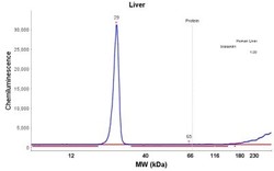
- Simple Western: Sigma-1 R/OPRS1 Antibody [NBP1-82479] - Simple Western lane view shows a specific band for SIGMAR1 in 0.2 mg/ml of Liver (left), RT-4 (right) lysate. This experiment was performed under reducing conditions using the 12-230 kDa separation system.

- Submitted by
- Novus Biologicals (provider)
- Main image

- Experimental details
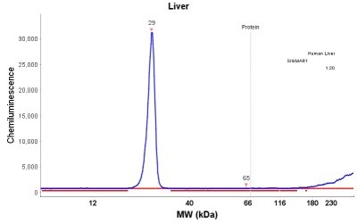
- Simple Western: Sigma-1 R/OPRS1 Antibody [NBP1-82479] - Electropherogram image(s) of corresponding Simple Western lane view. Sigma-1 R/OPRS1 antibody was used at 1:20 dilution on RT-4 lysate(s).

- Submitted by
- Novus Biologicals (provider)
- Main image
- Experimental details
- Simple Western: Sigma-1 R/OPRS1 Antibody [NBP1-82479] - Electropherogram image(s) of corresponding Simple Western lane view. Sigma-1 R/OPRS1 antibody was used at 1:20 dilution on Liver lysate(s).
- Submitted by
- Novus Biologicals (provider)
- Main image

- Experimental details
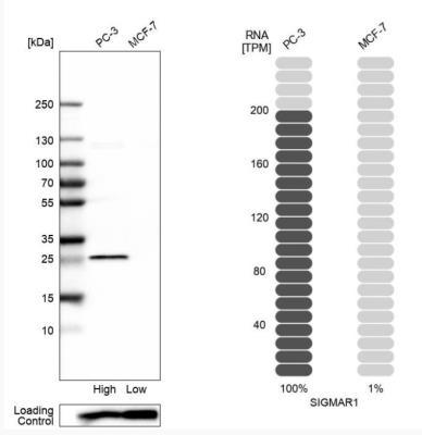
- Western Blot: Sigma-1 R/OPRS1 Antibody [NBP1-82479] - Analysis in human cell lines PC-3 and MCF-7 using Anti-SIGMAR1 antibody. Corresponding SIGMAR1 RNA-seq data are presented for the same cell lines. Loading control: Anti-PPIB.
Supportive validation
- Submitted by
- Novus Biologicals (provider)
- Main image
- Experimental details
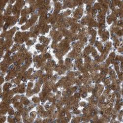
- Immunohistochemistry-Paraffin: Sigma-1 R/OPRS1 Antibody [NBP1-82479] - Staining of human liver shows strong cytoplasmic positivity in hepatocytes.

- Submitted by
- Novus Biologicals (provider)
- Main image
- Experimental details
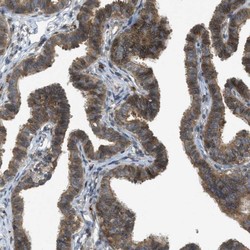
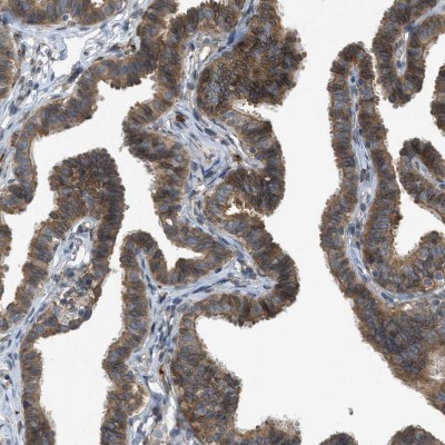
- Immunohistochemistry-Paraffin: Sigma-1 R/OPRS1 Antibody [NBP1-82479] - Staining of human Fallopian tube shows moderate cytoplasmic and membranous positivity in glandular cells.
- Submitted by
- Novus Biologicals (provider)
- Main image
- Experimental details
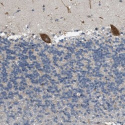
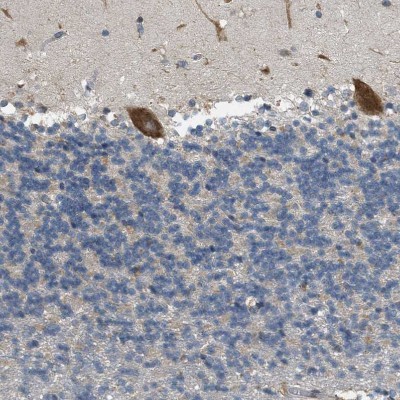
- Immunohistochemistry-Paraffin: Sigma-1 R/OPRS1 Antibody [NBP1-82479] - Staining of human cerebellum shows strong cytoplasmic positivity in Purkinje cells.
- Submitted by
- Novus Biologicals (provider)
- Main image

- Experimental details
- Immunohistochemistry-Paraffin: Sigma-1 R/OPRS1 Antibody [NBP1-82479] - Staining of human gastrointestinal shows moderate cytoplasmic and membranous positivity in glandular cells.
