 Explore
 Validate
 Learn
Learn Western blot
Antibody data
- Antibody Data
- Antigen structure
- References [1]
- Comments [0]
- Validations
- Western blot [1]
- Immunocytochemistry [1]
- Immunohistochemistry [8]
- Other assay [1]
Submit
Validation data
Reference
Comment
Report error
- Product number
- MA5-31383 - Provider product page

- Provider
- Invitrogen Antibodies
- Product name
- ChAT Monoclonal Antibody (CL3173)
- Antibody type
- Monoclonal
- Antigen
- Recombinant full-length protein
- Description
- Immunogen sequence: GLFSSYRLPG HTQDTLVAQN SSIMPEPEHV IVACCNQFFV LDVVINFRRL SEGDLFTQLR KIVKMASNED ERLPPIGLLT SDGRSEWAEA RTVLVKDSTN
- Reactivity
- Human, Mouse, Rat
- Host
- Mouse
- Isotype
- IgG
- Antibody clone number
- CL3173
- Vial size
- 100 µL
- Concentration
- 1 mg/mL
- Storage
- Store at 4°C short term. For long term storage, store at -20°C, avoiding freeze/thaw cycles.
Submitted references Involvement of neuronal and muscular Trk-fused gene (TFG) defects in the development of neurodegenerative diseases.
Yamamotoya T, Hasei S, Akasaka Y, Ohata Y, Nakatsu Y, Kanna M, Fujishiro M, Sakoda H, Ono H, Kushiyama A, Misawa H, Asano T
Scientific reports 2022 Feb 4;12(1):1966
Scientific reports 2022 Feb 4;12(1):1966
No comments: Submit comment
Supportive validation
- Submitted by
- Invitrogen Antibodies (provider)
- Main image

- Experimental details
- Western blot was performed using Anti-ChAT Monoclonal Antibody (CL3173) (Product # MA5-31383) and a ~70 kDa band corresponding to acetyl CoA: choline O-acetyltransferase was observed in Mouse and Rat Brain lysates but not in Mouse and Rat Kidney lysates. Whole cell extracts (30 µg lysate) of Mouse Brain (Lane 1), Rat Brain (Lane 2), Mouse Kidney (Lane 3) and Rat Kidney (Lane 4) were electrophoresed using NuPAGE™ 4-12% Bis-Tris Protein Gel (Product # NP0322BOX), 12 well. Resolved proteins were then transferred onto a nitrocellulose membrane (Product # IB23001) by iBlot® 2 Dry Blotting System (Product # IB21001). The blot was probed with the primary antibody (1:500 dilution) and detected by chemiluminescence with Goat anti-Mouse IgG (H+L) Superclonal™ Recombinant Secondary Antibody, HRP (Product # A28177, 1:20,000 dilution) using the iBright™ FL1500 Imaging System (Product # A44115). Chemiluminescent detection was performed using SuperSignal™ West Atto Ultimate Sensitivity Substrate (Product # A38556). Mouse Light chain band (*) was observed at ~25 kDa in Mouse Brain and Kidney lysates and an uncharacterized band (*) was observed in Rat Kidney lysates ~90 kDa.

Supportive validation
- Submitted by
- Invitrogen Antibodies (provider)
- Main image

- Experimental details
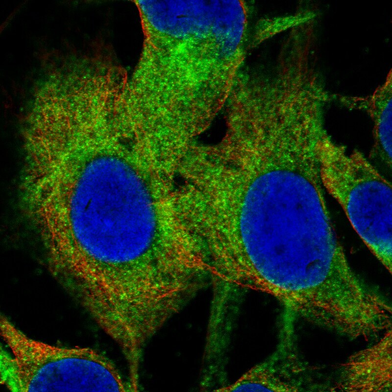
- Immunocytochemistry-Immunofluorescence analysis of ChAT in SK-MEL-30 cells using ChAT Monoclonal Antibody (CL3173) (Product # MA5-31383), showing specific staining in the cytosol in green. Microtubule- and nuclear probes are visualized in red and blue, respectively (where available).
Supportive validation
- Submitted by
- Invitrogen Antibodies (provider)
- Main image

- Experimental details
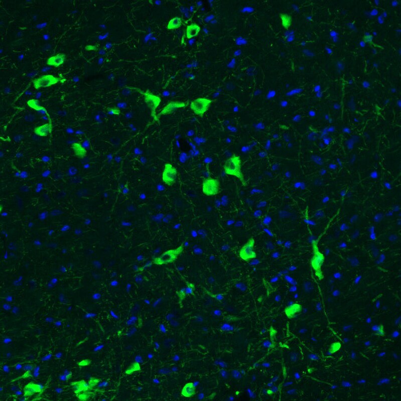
- Immunohistochemical analysis of ChAT in mouse basal forebrain using a ChAT monoclonal antibody (Product # MA5-31383). The analysis shows strong positivity in acetylcholine neurons in the caudate putamen.

- Submitted by
- Invitrogen Antibodies (provider)
- Main image

- Experimental details
- Immunohistochemical analysis of ChAT in rat brain using a ChAT monoclonal antibody (Product # MA5-31383). The analysis shows strong positivity in acetylcholine neurons in the basal forebrain.
- Submitted by
- Invitrogen Antibodies (provider)
- Main image

- Experimental details
- Immunohistochemical analysis of ChAT in mouse brain using a ChAT monoclonal antibody (Product # MA5-31383). The analysis shows strong positivity in acetylcholine neurons in the basal forebrain.

- Submitted by
- Invitrogen Antibodies (provider)
- Main image

- Experimental details
- Immunohistochemical analysis of ChAT in mouse basal forebrain using a ChAT monoclonal antibody (Product # MA5-31383). The analysis shows strong positivity in acetylcholine neurons in the caudate putamen.

- Submitted by
- Invitrogen Antibodies (provider)
- Main image

- Experimental details
- Immunohistochemical analysis of ChAT in human cerebral cortex using a ChAT monoclonal antibody (Product # MA5-31383). The analysis shows moderate positivity in cholinergic neural fibers.

- Submitted by
- Invitrogen Antibodies (provider)
- Main image

- Experimental details
- Immunohistochemical analysis of ChAT in human placenta using a ChAT monoclonal antibody (Product # MA5-31383). The analysis shows strong cytoplasmic positivity in a subset of cells in chorionic villi.

- Submitted by
- Invitrogen Antibodies (provider)
- Main image

- Experimental details
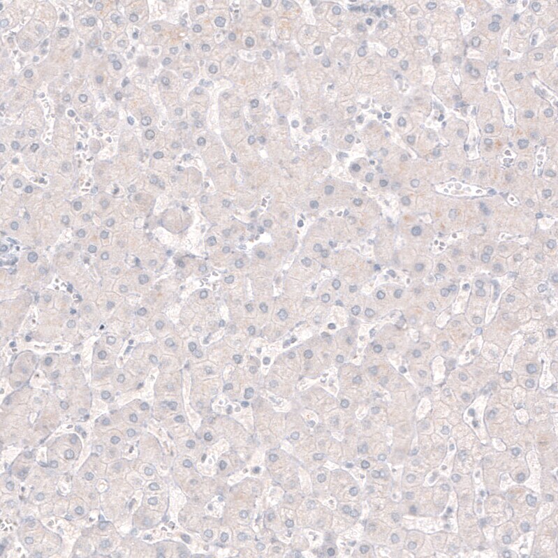
- Immunohistochemical analysis of ChAT in human liver using a ChAT monoclonal antibody (Product # MA5-31383). The analysis shows no positivity in hepatocytes as expected.
- Submitted by
- Invitrogen Antibodies (provider)
- Main image

- Experimental details
- Immunohistochemical analysis of ChAT in human placenta and liver tissues using a ChAT monoclonal antibody (Product # MA5-31383). Corresponding RNA-seq data are presented for the same tissues.

Supportive validation
- Submitted by
- Invitrogen Antibodies (provider)
- Main image

- Experimental details
- Figure 1 vMNTFG KO mice display reduced motor function (muscle weakness). ( a ) Fluorescent immunostaining against ChAT and TFG in lumbar spinal cord specimens from 6-month-old mice (scale bar: 100 um). ChAT-positive but TFG-negative motor neuron in vMNTFG KO is indicated by arrowhead. ( b ) Percentage of TFG-negative neurons among ChAT-positive motor neurons. ChAT-positive motor neurons number 213 and 177 from f/f (n = 6) and KO (n = 5), respectively. ( c ) Treadmill test results of 4-month-old male mice (n = 4). Distance (left panel) and time (right panel) that mice ran are shown. ( d ) Latency to fall in hanging-wire test results of 4-month-old male mice (n = 4-6). ( e ) Chronological changes in performance of the hanging-wire test (n = 8-9). (* P < 0.05, ** P < 0.01).
