Explore
 Validate
 Learn
Learn Western blot
Antibody data
- Antibody Data
- Antigen structure
- References [3]
- Comments [0]
- Validations
- Western blot [1]
- Immunohistochemistry [1]
Submit
Validation data
Reference
Comment
Report error
- Product number
- sc-259 - Provider product page

- Provider
- Santa Cruz Biotechnology
- Proper citation
- Santa Cruz Biotechnology Cat#sc-259, RRID:AB_2270724
- Product name
- Anti-SOS1
- Antibody type
- Polyclonal (Antigen purified)
- Antigen
- Synthetic peptide
- Reactivity
- Human
- Host
- Rabbit
Submitted references TGFbeta1 signaling via alphaVbeta6 integrin.
Ras-guanine nucleotide exchange factor sos2 is dispensable for mouse growth and development.
M2 alpha-1-antitrypsin phenotype and primary liver cancer
Kracklauer MP, Schmidt C, Sclabas GM
Molecular cancer 2003 Aug 7;2:28
Molecular cancer 2003 Aug 7;2:28
Ras-guanine nucleotide exchange factor sos2 is dispensable for mouse growth and development.
Esteban LM, Fernández-Medarde A, López E, Yienger K, Guerrero C, Ward JM, Tessarollo L, Santos E
Molecular and cellular biology 2000 Sep;20(17):6410-3
Molecular and cellular biology 2000 Sep;20(17):6410-3
M2 alpha-1-antitrypsin phenotype and primary liver cancer
P Sizaret, M Clerc, J Estève, R R Frants, J Pillot
British Journal of Cancer 1981 Feb;43(2):226-228
British Journal of Cancer 1981 Feb;43(2):226-228
No comments: Submit comment
Supportive validation
- Submitted by
- per
- Main image

- Experimental details
- Western blot analysis of antibody specificity using a routine panel composed of IgG/HSA-depleted human plasma and protein lysates from selected human tissues and cell lines.
- Validation comment
- No bands detected.
- Primary Ab dilution
- 1:500
- Secondary Ab dilution
- 1:3000
- Lane 1
- Marker [kDa]: 220, 112, 84, 47, 32, 26, 16.8
- Lane 2
- RT-4
- Lane 3
- U-251MG sp
- Lane 4
- Human Plasma
- Lane 5
- Liver
- Lane 6
- Tonsil
- Theoretical target weight
- [kDa] 128

Supportive validation
- Submitted by
- per
- Main image

- Experimental details
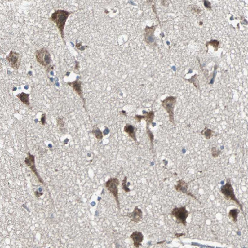
- Immunohistochemical staining of human cerebral cortex shows distinct cytoplasmic positivity in neuronal cells.
- Validation comment
- Staining pattern partly consistent with experimental and/or bioinformatic data.