Explore
 Validate
 Learn
LearnGTX25439
antibody from GeneTex
Targeting: HSPA1A
HSP70-1, HSPA1
 Western blot
 ELISA Immunocytochemistry Immunoprecipitation Immunohistochemistry Flow cytometry Blocking/Neutralizing Gel shift
Antibody data
- Antibody Data
- Antigen structure
- References [7]
- Comments [0]
- Validations
- Western blot [3]
- Immunocytochemistry [1]
- Immunoprecipitation [2]
Submit
Validation data
Reference
Comment
Report error
- Product number
- GTX25439 - Provider product page

- Provider
- GeneTex
- Proper citation
- GeneTex Cat#GTX25439, RRID:AB_373941
- Product name
- Hsp70 antibody [3A3]
- Antibody type
- Monoclonal
- Reactivity
- Human, Mouse, Rat, Chicken/Avian, Drosophila, Porcine, Simian, Yeast
- Host
- Mouse
Submitted references Identification of the gene encoding the TATA box-binding protein-associated factor 1 (TAF1) and its putative role in the heat shock response in the protozoan parasite Entamoeba histolytica.
Caveolin-1 Secreted from Adipose Tissues and Adipocytes Functions as an Adipogenesis Enhancer.
Extracellular Hsp70 Enhances Mesoangioblast Migration via an Autocrine Signaling Pathway.
HSP70 regulates the function of mitotic centrosomes.
Immunohistochemical distribution of heat shock protein 70 and proliferating cell nuclear antigen in mouse placenta at different gestational stages.
A calreticulin-dependent nuclear export signal is involved in the regulation of liver receptor homologue-1 protein folding.
Hyperbaric oxygen preconditioning induces tolerance against oxidative injury and oxygen-glucose deprivation by up-regulating heat shock protein 32 in rat spinal neurons.
Avendaño-Borromeo B, Narayanasamy RK, García-Rivera G, Labra-Barrios ML, Lagunes-Guillén AE, Munguía-Chávez B, Castañón-Sánchez CA, Orozco E, Luna-Arias JP
Parasitology research 2019 Feb;118(2):517-538
Parasitology research 2019 Feb;118(2):517-538
Caveolin-1 Secreted from Adipose Tissues and Adipocytes Functions as an Adipogenesis Enhancer.
Chang CC, Chen CY, Wen HC, Huang CY, Hung MS, Lu HC, Chen WL, Chang CH
Obesity (Silver Spring, Md.) 2017 Nov;25(11):1932-1940
Obesity (Silver Spring, Md.) 2017 Nov;25(11):1932-1940
Extracellular Hsp70 Enhances Mesoangioblast Migration via an Autocrine Signaling Pathway.
Barreca MM, Spinello W, Cavalieri V, Turturici G, Sconzo G, Kaur P, Tinnirello R, Asea AA, Geraci F
Journal of cellular physiology 2017 Jul;232(7):1845-1861
Journal of cellular physiology 2017 Jul;232(7):1845-1861
HSP70 regulates the function of mitotic centrosomes.
Fang CT, Kuo HH, Pan TS, Yu FC, Yih LH
Cellular and molecular life sciences : CMLS 2016 Oct;73(20):3949-60
Cellular and molecular life sciences : CMLS 2016 Oct;73(20):3949-60
Immunohistochemical distribution of heat shock protein 70 and proliferating cell nuclear antigen in mouse placenta at different gestational stages.
Ozaydin T, Sur E, Oznurlu Y, Celik I, Uluisik D
Microscopy research and technique 2016 Apr;79(4):251-7
Microscopy research and technique 2016 Apr;79(4):251-7
A calreticulin-dependent nuclear export signal is involved in the regulation of liver receptor homologue-1 protein folding.
Yang FM, Feng SJ, Lai TC, Hu MC
The Biochemical journal 2015 Oct 15;471(2):199-209
The Biochemical journal 2015 Oct 15;471(2):199-209
Hyperbaric oxygen preconditioning induces tolerance against oxidative injury and oxygen-glucose deprivation by up-regulating heat shock protein 32 in rat spinal neurons.
Huang G, Xu J, Xu L, Wang S, Li R, Liu K, Zheng J, Cai Z, Zhang K, Luo Y, Xu W
PloS one 2014;9(1):e85967
PloS one 2014;9(1):e85967
No comments: Submit comment
Supportive validation
- Submitted by
- GeneTex (provider)
- Main image

- Experimental details
- Western blot analysis of HSP70 in 50μg of various cell lysates. Proteins were transferred to a PVDF membrane and blocked with 5% BSA/TBST for at least 1 hour. The membrane was probed with HSP70 antibody [3A3] at a dilution of 1:1000 overnight at 4°C on a rocking platform, washed in TBS-0.1%Tween 20, and probed with a proper secondary antibody. Chemiluminescent detection was performed.

- Submitted by
- GeneTex (provider)
- Main image

- Experimental details
- Immunofluorescent analysis of Heat Shock Protein 70 using Heat Shock Protein 70 Monoclonal antibody (3A3) (GTX25439) shows staining in NIH-3T3 cells. Heat Shock Protein 70 staining (green), F-Actin staining with Phalloidin (red) and nuclei with DAPI (blue) is shown. Cells were grown on chamber slides and fixed with formaldehyde prior to staining. The antibody dilution is 1:100-1:200.
- Validation comment
- WB
- Submitted by
- GeneTex (provider)
- Main image

- Experimental details
- Immunofluorescent analysis of Heat Shock Protein 70 using Heat Shock Protein 70 Monoclonal antibody shows staining in MCF-7 cells. Heat Shock Protein 70 staining (green), F-Actin staining with Phalloidin (red) and nuclei with DAPI (blue) is shown. Cells were grown on chamber slides and fixed with formaldehyde prior to staining. Cells were probed without (control) or with or an antibody recognizing Heat Shock Protein 70 (GTX25439) at a dilution of 1:100-1:200 over night at 4 °C, washed with PBS and incubated with a DyLight-488 conjugated secondary antibody. Images were taken at 60X magnification.
- Validation comment
- WB

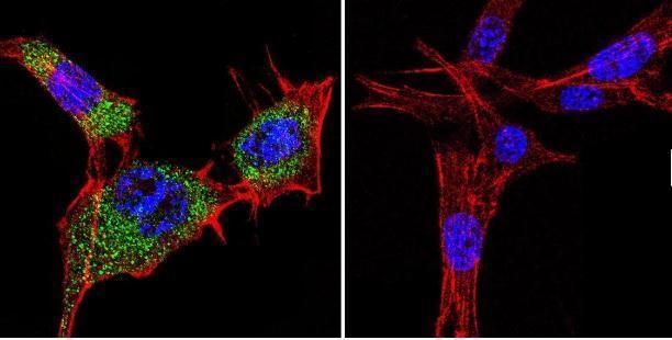
Supportive validation
- Submitted by
- GeneTex (provider)
- Main image

- Experimental details
- Immunofluorescent analysis of HSP70 in NIH-3T3 cells. HSP70 staining (green), F-Actin staining with Phalloidin (red) and nuclei with DAPI (blue) is shown. Cells were grown on slides and fixed with formaldehyde prior to staining. Cells were probed without (control) or with HSP70 antibody [3A3] at a dilution of 1:100-1:200 over night at 4°C, washed with PBS and incubated with a proper secondary antibody. Images were taken at 60X magnification.

Supportive validation
- Submitted by
- GeneTex (provider)
- Main image

- Experimental details
- Immunoprecipitation of HSP70 in HeLa cells. Antigen-antibody complexes were formed by incubating 500ug whole cell lysate with 2ul of HSP70 antibody [3A3] overnight on a rocking platform at 4¢XC. The immune complexes were captured on 50ul Protein A/G Agarose , washed extensively, and eluted. Samples were then resolved on a 4-20% Tris-HCl polyacrylamide gel, transferred to a PVDF membrane, and blocked with 5% BSA/TBST for at least 1 hour. The membrane was probed with HSP70 antibody [3A3] at a dilution of 1:1000 overnight rotating at 4¢XC, washed in TBST, and probed with a proper secondary antibody. Chemiluminescent detection was performed.

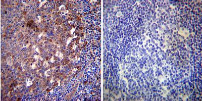
- Submitted by
- GeneTex (provider)
- Main image

- Experimental details
- Immunohistochemistry was performed on normal deparaffinized human Tonsil tissue. To expose target proteins, heat induced antigen retrieval was performed using 10mM sodium citrate (pH 6.0) buffer, microwaved for 8-15 minutes. Following antigen retrieval tissues were blocked in 3% BSA-PBS for 30 minutes at room temperature. Tissues were then probed at a dilution of 1:20 with a mouse monoclonal antibody recognizing Heat Shock Protein 70 (GTX25439) or without primary antibody (negative control) overnight at 4°C in a humidified chamber. Tissues were washed extensively with PBST and endogenous peroxidase activity was quenched with a peroxidase suppressor. Detection was performed using a biotin-conjugated secondary antibody and SA-HRP, followed by colorimetric detection using DAB. Tissues were counterstained with hematoxylin and prepped for mounting.
- Validation comment
- IP