 Explore
 Validate
 Learn
LearnGTX22787
antibody from GeneTex
Targeting: HSPA1A
HSP70-1, HSPA1
 Western blot
 ELISA Immunocytochemistry Immunoprecipitation Immunohistochemistry Blocking/Neutralizing Gel shift
Antibody data
- Antibody Data
- Antigen structure
- References [0]
- Comments [0]
- Validations
- Western blot [1]
- Immunocytochemistry [1]
- Immunoprecipitation [1]
Submit
Validation data
Reference
Comment
Report error
- Product number
- GTX22787 - Provider product page

- Provider
- GeneTex
- Proper citation
- GeneTex Cat#GTX22787, RRID:AB_384850
- Product name
- Hsp70 antibody [5A5]
- Antibody type
- Monoclonal
- Reactivity
- Human, Mouse, Rat, Bovine, Chicken/Avian, Drosophila, Rabbit, Yeast
- Host
- Mouse
No comments: Submit comment
Supportive validation
- Submitted by
- GeneTex (provider)
- Main image

- Experimental details
- Western blot analysis of HSP70 in 50μg of various cell lysates. Proteins were transferred to a PVDF membrane and blocked with 5% BSA/TBST for at least 1 hour. The membrane was probed with HSP70 antibody [5A5] at a dilution of 1:1000 overnight at 4°C on a rocking platform, washed in TBS-0.1%Tween 20, and probed with a proper secondary antibody. Chemiluminescent detection was performed.

Supportive validation
- Submitted by
- GeneTex (provider)
- Main image

- Experimental details
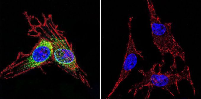
- Immunofluorescent analysis of HSP70 in C6 glioma cells. HSP70 staining (green), F-Actin staining with Phalloidin (red) and nuclei with DAPI (blue) is shown. Cells were grown on slides and fixed with formaldehyde prior to staining. Cells were probed without (control) or with HSP70 antibody [5A5] at a dilution of 1:100-1:200 over night at 4 °C, washed with PBS and incubated with a proper secondary antibody. Imges were taken at 60X magnification.
Supportive validation
- Submitted by
- GeneTex (provider)
- Main image

- Experimental details
- Immunoprecipitation of HSP70 in HeLa cells. Antigen-antibody complexes were formed by incubating 500£gg whole cell lysate with 2£gg of HSP70 monoclonal antibody overnight on a rocking platform at 4¢XC. The immune complexes were captured on 50£gl Protein A/G Agarose , washed extensively, and eluted. Samples were then resolved on a 4-20% Tris-HCl polyacrylamide gel, transferred to a PVDF membrane, and blocked with 5% BSA/TBST for at least 1 hour. The membrane was probed with HSP70 antibody [5A5] at a dilution of 1:1000 overnight rotating at 4¢XC, washed in TBST, and probed with a proper secondary antibody. Chemiluminescent detection was performed.
