 Explore
 Validate
 Learn
Learn Western blot
Antibody data
- Antibody Data
- Antigen structure
- References [1]
- Comments [0]
- Validations
- Western blot [2]
- Immunocytochemistry [1]
- Immunohistochemistry [2]
- Flow cytometry [1]
Submit
Validation data
Reference
Comment
Report error
- Product number
- TA500772 - Provider product page

- Provider
- OriGene
- Proper citation
- OriGene Cat#TA500772, RRID:AB_11125504
- Product name
- Mouse monoclonal anti-HSPA1A(HSP70) antibody, clone 3C6, Loading, clone OTI3C6 (formerly 3C6)
- Antibody type
- Monoclonal
- Description
- Mouse monoclonal anti-HSPA1A(HSP70) antibody, clone 3C6, Loading, clone OTI3C6 (formerly 3C6)
- Reactivity
- Canine
- Host
- Mouse
- Conjugate
- Unconjugated
- Epitope
- HSPA1A
- Isotype
- IgG
- Antibody clone number
- OTI3C6
- Vial size
- 100 µl
- Concentration
- NULL
Submitted references Tracking protein aggregation and mislocalization in cells with flow cytometry.
Ramdzan YM, Polling S, Chia CP, Ng IH, Ormsby AR, Croft NP, Purcell AW, Bogoyevitch MA, Ng DC, Gleeson PA, Hatters DM
Nature methods 2012 Mar 18;9(5):467-70
Nature methods 2012 Mar 18;9(5):467-70
No comments: Submit comment
Supportive validation
- Submitted by
- OriGene (provider)
- Main image

- Experimental details
- Western blot analysis of extracts (35ug) from 9 different cell lines by using anti-HSPA1A monoclonal antibody.
- Validation comment
- WB

- Submitted by
- OriGene (provider)
- Main image
- Experimental details
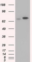
- HEK293T cells were transfected with the pCMV6-ENTRY control (Left lane) or pCMV6-ENTRY HSPA1A (RC200270, Right lane) cDNA for 48 hrs and lysed. Equivalent amounts of cell lysates (5 ug per lane) were separated by SDS-PAGE and immunoblotted with anti-HSPA1A.
- Validation comment
- WB

Supportive validation
- Submitted by
- OriGene (provider)
- Main image
- Experimental details
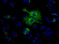
- Anti-HSPA1A mouse monoclonal antibody (TA500772) immunofluorescent staining of COS7 cells transiently transfected by pCMV6-ENTRY HSPA1A(RC200270).
- Validation comment
- IF

Supportive validation
- Submitted by
- OriGene (provider)
- Main image
- Experimental details
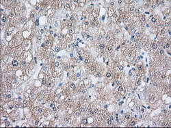
- Immunohistochemical staining of paraffin-embedded Human liver tissue within the normal limits using anti-HSPA1A mouse monoclonal antibody. (Heat-induced epitope retrieval by 10mM citric buffer, pH6.0, 100C for 10min, TA500772, Dilution 1:50)
- Validation comment
- IHC

- Submitted by
- OriGene (provider)
- Main image
- Experimental details
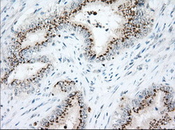
- Immunohistochemical staining of paraffin-embedded Adenocarcinoma of Human colon tissue using anti-HSPA1A mouse monoclonal antibody. (Heat-induced epitope retrieval by 10mM citric buffer, pH6.0, 100C for 10min, TA500772, Dilution 1:50)
- Validation comment
- IHC

Supportive validation
- Submitted by
- OriGene (provider)
- Main image
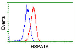
- Experimental details
- Flow cytometric Analysis of Hela cells, using anti-HSPA1A antibody(TA500772),(Red), compared to a nonspecific negative control antibody(TA50011),(Blue).
- Validation comment
- FC
