 Explore
 Validate
 Learn
Learn Western blot
Antibody data
- Antibody Data
- Antigen structure
- References [2]
- Comments [0]
- Validations
- Western blot [2]
- Immunohistochemistry [1]
Submit
Validation data
Reference
Comment
Report error
- Product number
- MAB1663 - Provider product page

- Provider
- R&D Systems
- Product name
- Human/Mouse/Rat HSP70/HSPA1A Antibody
- Antibody type
- Monoclonal
- Description
- Protein A or G purified from hybridoma culture supernatant. Detects the induced form of human and mouse HSP70/HSPA1A in Western blots. In Western blots, no cross-reactivity with the constitutively expressed HSC70 (HSP73) is detected.
- Reactivity
- Human, Mouse, Rat
- Host
- Mouse
- Conjugate
- Unconjugated
- Antigen sequence
NP_005336- Isotype
- IgG
- Antibody clone number
- 242707
- Vial size
- 100 ug
- Concentration
- LYOPH
- Storage
- Use a manual defrost freezer and avoid repeated freeze-thaw cycles. 12 months from date of receipt, -20 to -70 °C as supplied. 1 month, 2 to 8 °C under sterile conditions after reconstitution. 6 months, -20 to -70 °C under sterile conditions after reconstitution.
Submitted references Comparative analysis of the interaction of HSPs in dendritic cells, macrophages, RGM-1 cells infected by Helicobacter pylori.
Exosomes released from macrophages infected with intracellular pathogens stimulate a proinflammatory response in vitro and in vivo.
Yao Y, Wu J, Gu T, Cheng Y, Li G
American journal of translational research 2016;8(10):4184-4194
American journal of translational research 2016;8(10):4184-4194
Exosomes released from macrophages infected with intracellular pathogens stimulate a proinflammatory response in vitro and in vivo.
Bhatnagar S, Shinagawa K, Castellino FJ, Schorey JS
Blood 2007 Nov 1;110(9):3234-44
Blood 2007 Nov 1;110(9):3234-44
No comments: Submit comment
Supportive validation
- Submitted by
- R&D Systems (provider)
- Main image
- Experimental details
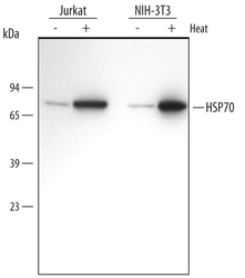
- Detection of Human and Mouse HSP70/ HSPA1A by Western Blot. Western blot shows lysates of Jurkat human acute T cell leukemia cell line and NIH-3T3 mouse embryonic fibroblast cell line untreated (-) or treated (+) with a 42 °C heat shock for 30 minutes with a 3 hour recovery. PVDF membrane was probed with 0.1 µg/mL of Mouse Anti-Human/Mouse/Rat HSP70/HSPA1A Monoclonal Antibody (Catalog # MAB1663), followed by HRP-conjugated Anti-Mouse IgG Secondary Antibody (Catalog # HAF007). A specific band was detected for HSP70/HSPA1A at approximately 70 kDa (as indicated). This experiment was conducted under reducing conditions and using Immunoblot Buffer Group 2.
- Submitted by
- R&D Systems (provider)
- Main image
- Experimental details
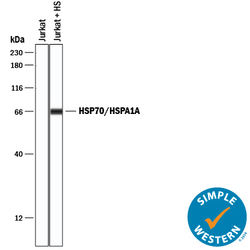
- Detection of Human HSP70/HSPA1A by Simple WesternTM. Simple Western lane view shows lysates of Jurkat human acute T cell leukemia cell line untreated (-) or treated (+) by heat shocked (HS), loaded at 0.2 mg/mL. A specific band was detected for HSP70/HSPA1A at approximately 67 kDa (as indicated) using 0.5 µg/mL of Mouse Anti-Human/Mouse/Rat HSP70/HSPA1A Monoclonal Antibody (Catalog # MAB1663) . This experiment was conducted under reducing conditions and using the 12-230 kDa separation system.

Supportive validation
- Submitted by
- R&D Systems (provider)
- Main image
- Experimental details
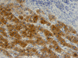
- HSP70/HSPA1A in Human Liver Cancer Tissue. HSP70/HSPA1A was detected in immersion fixed paraffin-embedded sections of human liver cancer tissue using Mouse Anti-Human/Mouse/Rat HSP70/HSPA1A Monoclonal Antibody (Catalog # MAB1663) at 25 µg/mL overnight at 4 °C. Tissue was stained using the Anti-Mouse HRP-DAB Cell & Tissue Staining Kit (brown; Catalog # CTS002) and counterstained with hematoxylin (blue). View our protocol for Chromogenic IHC Staining of Paraffin-embedded Tissue Sections.
