 Explore
 Validate
 Learn
Learn Immunocytochemistry
 Immunohistochemistry
Antibody data
- Antibody Data
- Antigen structure
- References [1]
- Comments [0]
- Validations
- Immunohistochemistry [1]
Submit
Validation data
Reference
Comment
Report error
- Product number
- NB100-65219 - Provider product page

- Provider
- Novus Biologicals
- Proper citation
- Novus Cat#NB100-65219, RRID:AB_965531
- Product name
- Rat Monoclonal Substance P Antibody
- Antibody type
- Monoclonal
- Description
- Tissue culture supernatant. NB100-65219 recognizes the COOH terminal end of substance P, a short polypeptide neurotransmitter that regulates the excitability of dorsal horn nociceptive neurons. It is known to have a role in several physiologic activities such as pain transmission, the vomiting reflex, salivary secretion and smooth muscle contraction. 5% reactivity is observed with eledoisin. It does not react with Leu or Met-enkephalin, somatostatin or beta-endorphin.
- Reactivity
- Human, Mouse
- Host
- Rat
- Isotype
- IgG
- Vial size
- 0.05 ml
- Storage
- Store at 4C short term. Aliquot and store at -20C long term. Avoid freeze-thaw cycles.
Submitted references Localisation and activation of the neurokinin 1 receptor in the enteric nervous system of the mouse distal colon.
Pelayo JC, Veldhuis NA, Eriksson EM, Bunnett NW, Poole DP
Cell and tissue research 2014 May;356(2):319-32
Cell and tissue research 2014 May;356(2):319-32
No comments: Submit comment
Supportive validation
- Submitted by
- Novus Biologicals (provider)
- Main image
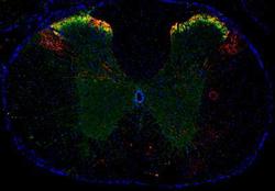
- Experimental details
- Immunohistochemistry-Frozen: Substance P Antibody (NC1/34) [NB100-65219] - Opioid R/OPRM1 (green) and Substance P (red) were detected in frozen mouse spinal cord and DRG tissue by fluorescent multiplex IHC. Mu Opioid R/OPRM1 Antibody (NB100-1620), used at 1:100, and Substance P Antibody (NB100-65219), used at 1:50, were applied to the tissue and simultaneously incubated at 4C overnight. Anti-Rabbit Alexa Fluor(R) 488-conjugated secondary antibody and anti-Rat NL557-conjugated secondary antibody (NL013) were applied to the tissue, and simultaneously incubated at room temperature, protected from light for 1 hour. Coverslips were applied to the tissue slide along with mounting media containing DAPI (blue) and imaged using a 4X objective lens.
