 Explore
 Validate
 Learn
Learn Immunocytochemistry
Antibody data
- Antibody Data
- Antigen structure
- References [2]
- Comments [0]
- Validations
- Immunocytochemistry [1]
- Immunohistochemistry [1]
Submit
Validation data
Reference
Comment
Report error
- Product number
- HPA001619 - Provider product page

- Provider
- Atlas Antibodies
- Proper citation
- Atlas Antibodies Cat#HPA001619, RRID:AB_1078727
- Product name
- Anti-EIF4ENIF1
- Antibody type
- Polyclonal
- Reactivity
- Human
- Host
- Rabbit
- Conjugate
- Unconjugated
- Antigen sequence
FAPIPLEDHAENKVDILEMLQKAKVDLKPLLSSLS
ANKEKLKESSHSGVVLSVEEVEAGLKGLKVDQQVK
NSTPFMAEHLEETLSAVTNNRQLKKDGDMTAFNKL
VSTMKASGTLPSQPKVSRNLESHLMSPAEI- Isotype
- IgG
- Vial size
- 100 µl
- Storage
- Store at +4°C for short term storage. Long time storage is recommended at -20°C.
Submitted references Phosphorylation of eIF4E Confers Resistance to Cellular Stress and DNA-Damaging Agents through an Interaction with 4E-T: A Rationale for Novel Therapeutic Approaches.
Systematic validation of antibody binding and protein subcellular localization using siRNA and confocal microscopy
Martínez A, Sesé M, Losa JH, Robichaud N, Sonenberg N, Aasen T, Ramón Y Cajal S
PloS one 2015;10(4):e0123352
PloS one 2015;10(4):e0123352
Systematic validation of antibody binding and protein subcellular localization using siRNA and confocal microscopy
Stadler C, Hjelmare M, Neumann B, Jonasson K, Pepperkok R, Uhlén M, Lundberg E
Journal of Proteomics 2012 April;75(7):2236-2251
Journal of Proteomics 2012 April;75(7):2236-2251
No comments: Submit comment
Supportive validation
- Submitted by
- Atlas Antibodies (provider)
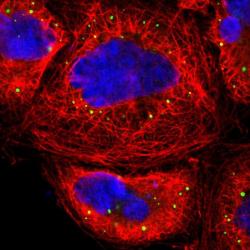
- Main image
- Experimental details
- Immunofluorescent staining of human cell line A-431 shows localization to vesicles.
- Sample type
- HUMAN

Supportive validation
- Submitted by
- Atlas Antibodies (provider)
- Main image

- Experimental details
- Immunohistochemical staining of human pancreas shows strong cytoplasmic positivity in exocrine glandular cells.
- Sample type
- HUMAN
