 Explore
 Validate
 Learn
Learn Western blot
Antibody data
- Antibody Data
- Antigen structure
- References [3]
- Comments [0]
- Validations
- Western blot [4]
- Immunohistochemistry [1]
Submit
Validation data
Reference
Comment
Report error
- Product number
- GTX114881 - Provider product page

- Provider
- GeneTex
- Proper citation
- GeneTex Cat#GTX114881, RRID:AB_11172941
- Product name
- Wnt7b antibody
- Antibody type
- Polyclonal
- Reactivity
- Human, Mouse
- Host
- Rabbit
Submitted references FGF23 Regulates Wnt/β-Catenin Signaling-Mediated Osteoarthritis in Mice Overexpressing High-Molecular-Weight FGF2.
Involvement of RARRES3 in the regulation of Wnt proteins acylation and signaling activities in human breast cancer cells.
Downregulated miR329 and miR410 promote the proliferation and invasion of oral squamous cell carcinoma by targeting Wnt-7b.
Meo Burt P, Xiao L, Hurley MM
Endocrinology 2018 Jun 1;159(6):2386-2396
Endocrinology 2018 Jun 1;159(6):2386-2396
Involvement of RARRES3 in the regulation of Wnt proteins acylation and signaling activities in human breast cancer cells.
Hsu TH, Jiang SY, Chang WL, Eckert RL, Scharadin TM, Chang TC
Cell death and differentiation 2015 May;22(5):801-14
Cell death and differentiation 2015 May;22(5):801-14
Downregulated miR329 and miR410 promote the proliferation and invasion of oral squamous cell carcinoma by targeting Wnt-7b.
Shiah SG, Hsiao JR, Chang WM, Chen YW, Jin YT, Wong TY, Huang JS, Tsai ST, Hsu YM, Chou ST, Yen YC, Jiang SS, Shieh YS, Chang IS, Hsiao M, Chang JY
Cancer research 2014 Dec 15;74(24):7560-72
Cancer research 2014 Dec 15;74(24):7560-72
No comments: Submit comment
Supportive validation
- Submitted by
- GeneTex (provider)
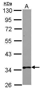
- Main image

- Experimental details
- Sample (30 ?g of whole cell lysate) A: NT2D1 10% SDS PAGE GTX114881 diluted at 1:1000 The HRP-conjugated anti-rabbit IgG antibody (GTX213110-01) was used to detect the primary antibody.
- Submitted by
- GeneTex (provider)
- Main image

- Experimental details
- Sample (30 ?g of whole cell lysate) A: Non-transfected 293T lysates B: WNT7B protein transfected 293T lysates 10% SDS PAGE GTX114881 diluted at 1:5000 The HRP-conjugated anti-rabbit IgG antibody (GTX213110-01) was used to detect the primary antibody.

- Submitted by
- GeneTex (provider)
- Main image

- Experimental details
- Wnt7b antibody detects Wnt7b protein by western blot analysis. Mouse tissue extracts (50 ?g) was separated by 10% SDS-PAGE, and the membrane was blotted with Wnt7b antibody (GTX114881) at a dilution of 1:1000. The HRP-conjugated anti-rabbit IgG antibody (GTX213110-01) was used to detect the primary antibody.

- Submitted by
- GeneTex (provider)
- Main image

- Experimental details
- Wnt7b antibody detects Wnt7b protein by western blot analysis. Non-transfected (-) and Wnt7b-transfected (+, including V5-tag) 293T whole cell extracts (72 ?g) were separated by 10% SDS-PAGE, and the membrane was blotted with Wnt7b antibody (GTX114881) at a dilution of 1:5000.The V5 was used as positive control (GTX117997, 1:2500) shown at the bottom panel. The HRP-conjugated anti-rabbit IgG antibody (GTX213110-01) was used to detect the primary antibody.

Supportive validation
- Submitted by
- GeneTex (provider)
- Main image

- Experimental details
- Immunohistochemical analysis of paraffin-embedded human ovarian carcinoma, using Wnt7b(GTX114881) antibody at 1:250 dilution.
