Explore
 Validate
 Learn
Learn Western blot
 ELISA
ELISAAntibody data
- Antibody Data
- Antigen structure
- References [1]
- Comments [0]
- Validations
- Western blot [1]
- Immunocytochemistry [2]
- Immunohistochemistry [1]
Submit
Validation data
Reference
Comment
Report error
- Product number
- AP16208PU-N - Provider product page

- Provider
- Acris Antibodies GmbH
- Proper citation
- Acris Antibodies GmbH Cat#AP16208PU-N, RRID:AB_1928175
- Product name
- anti VPS35 / MEM3 (C-term)
- Antibody type
- Polyclonal
- Antigen
- Syntehtic peptide from the C-Terminus of Human VPS35 (NP_060676.2).
- Reactivity
- Human, Mouse, Rat, Bovine
- Host
- Goat
- Vial size
- 0.1 mg
- Concentration
- 0.5 mg/ml
Submitted references Impaired retrograde membrane traffic through endosomes in a mutant CHO cell defective in phosphatidylserine synthesis.
Lee S, Uchida Y, Emoto K, Umeda M, Kuge O, Taguchi T, Arai H
Genes to cells : devoted to molecular & cellular mechanisms 2012 Aug;17(8):728-36
Genes to cells : devoted to molecular & cellular mechanisms 2012 Aug;17(8):728-36
No comments: Submit comment
Supportive validation
- Submitted by
- Acris Antibodies GmbH (provider)
- Main image

- Experimental details
- AP16208PU-N staining (0.05 µg/ml) of Human Brain Cerebellum lysate (RIPA buffer, 35µg total protein per lane). Primary incubated for 1 hour. Detected by western blot using chemiluminescence.

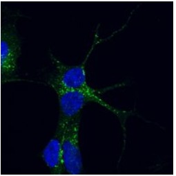
Supportive validation
- Submitted by
- Acris Antibodies GmbH (provider)
- Main image

- Experimental details
- AP16208PU-N staining (0.05 µg/ml) of PFA-fixed and saponin permeabilized SHSY5Y and detected with FITC in confocal microscopy. Data obtained from Dr. M. Schallburg Nielsen, Aarhus University Denmark.
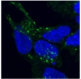
- Submitted by
- Acris Antibodies GmbH (provider)
- Main image

- Experimental details
- AP16208PU-N staining (0.05 µg/ml) of PFA-fixed and saponin permeabilized HEK293 and detected with FITC in confocal microscopy. Data obtained from Aarhus University Denmark.
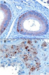
Supportive validation
- Submitted by
- Acris Antibodies GmbH (provider)
- Main image
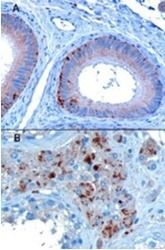
- Experimental details
- AP16208PU-N staining of paraffin embedded Human Testis showing A) Epithelial cells of the epididymis and B) Some leydig cells. Microwaved antigen retrieval with A) Tris/EDTA buffer pH9 at 3µg/ml or B) Citrate buffer pH6 at 10 µg/ml. HRP-staining.