Explore
 Validate
 Learn
Learn Western blot
 Immunoprecipitation
Antibody data
- Antibody Data
- Antigen structure
- References [1]
- Comments [0]
- Validations
- Western blot [8]
- Immunocytochemistry [3]
- Immunohistochemistry [1]
- Other assay [1]
Submit
Validation data
Reference
Comment
Report error
- Product number
- PA5-21898 - Provider product page

- Provider
- Invitrogen Antibodies
- Product name
- VPS35 Polyclonal Antibody
- Antibody type
- Polyclonal
- Antigen
- Synthetic peptide
- Description
- Recommended positive controls: Mouse brain, Rat Brain, Hela, A549, H1299, HCT116, HepG2, Molt4, Raji. Predicted reactivity: Mouse (100%), Rat (100%), Bovine (100%). Store product as a concentrated solution. Centrifuge briefly prior to opening the vial.
- Reactivity
- Human, Mouse, Rat
- Host
- Rabbit
- Isotype
- IgG
- Vial size
- 100 µL
- Concentration
- 0.66 mg/mL
- Storage
- Store at 4°C short term. For long term storage, store at -20°C, avoiding freeze/thaw cycles.
Submitted references Dopamine Transporter Localization in Medial Forebrain Bundle Axons Indicates Its Long-Range Transport Primarily by Membrane Diffusion with a Limited Contribution of Vesicular Traffic on Retromer-Positive Compartments.
Bagalkot TR, Block ER, Bucchin K, Balcita-Pedicino JJ, Calderon M, Sesack SR, Sorkin A
The Journal of neuroscience : the official journal of the Society for Neuroscience 2021 Jan 13;41(2):234-250
The Journal of neuroscience : the official journal of the Society for Neuroscience 2021 Jan 13;41(2):234-250
No comments: Submit comment
Supportive validation
- Submitted by
- Invitrogen Antibodies (provider)
- Main image
- Experimental details
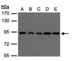
- Western blot analysis of VPS35 using 30 µg of A) H1299 (B) HeLa S3 (C) HepG2 (D) MOLT4 and E) Raji lysate. Samples were loaded onto a 7.5% SDS-PAGE gel and probed with a VPS35 polyclonal antibody (Product # PA5-21898) at a dilution of 1:500.

- Submitted by
- Invitrogen Antibodies (provider)
- Main image

- Experimental details
- Western blot analysis of VPS35 in A) 30 µg A549 whole cell extract, B) 30 µg H1299 whole cell extract, and C) 30 µg HCT116 whole cell extract. Samples were separated by 7.5% SDS-PAGEand the membrane was probed with VPS35, C-term Polyclonal antibody (Product # PA5-21898) at a dilution of 1:1000.

- Submitted by
- Invitrogen Antibodies (provider)
- Main image

- Experimental details
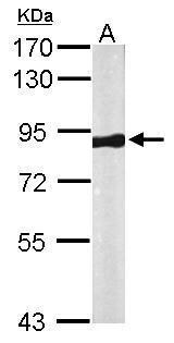
- Western Blot using VPS35 Polyclonal Antibody (Product # PA5-21898). Sample (50 µg of whole cell lysate). Lane A: Mouse brain. 7.5% SDS PAGE. VPS35 Polyclonal Antibody (Product # PA5-21898) diluted at 1:1,000. The HRP-conjugated anti-rabbit IgG antibody was used to detect the primary antibody.
- Submitted by
- Invitrogen Antibodies (provider)
- Main image

- Experimental details
- Western Blot using VPS35 Polyclonal Antibody (Product # PA5-21898). Various whole cell extracts (30 µg) were separated by 7.5% SDS-PAGE, and the membrane was blotted with VPS35 Polyclonal Antibody (Product # PA5-21898) diluted at 1:1,000. The HRP-conjugated anti-rabbit IgG antibody was used to detect the primary antibody.

- Submitted by
- Invitrogen Antibodies (provider)
- Main image

- Experimental details
- VPS35 Polyclonal Antibody detects VPS35 protein by western blot analysis. A. 50 µg rat brain lysate/extract.7.5% SDS-PAGE. VPS35 Polyclonal Antibody (Product # PA5-21898) dilution: 1:500. The HRP-conjugated anti-rabbit IgG antibody was used to detect the primary antibody.

- Submitted by
- Invitrogen Antibodies (provider)
- Main image

- Experimental details
- Western blot of VPS35 was performed by loading 20 µg of HAP1 WT (lane 1) and VPS35 KO (lane 2) cell lysates in RIPA buffer onto a 4-15% gradient polyacrylamide gel. Proteins on the blot were visualized with Ponceau staining (below immunoblot). Proteins were transferred to nitrocellulose membrane and blocked in 5% milk for 1 hr. VPS35 was detected at approximately 91 kDa using VPS35 Polyclonal Antibody (Product # PA5-21898) at a dilution of 1:1000 in 5% BSA in TBST overnight at 4 degree celsius. The blot was probed with Goat anti-Rabbit IgG (H+L) Secondary Antibody, HRP (Product # 65-6120) diluted to 0.2 µg/mL in TBST with 5% milk for 1 hr at room temperature. Chemiluminescent detection was performed using ECL Western Blotting Substrate. Data courtesy of YCharOS Inc., an open science company with the mission of characterizing commercially available antibodies using knockout validation.

- Submitted by
- Invitrogen Antibodies (provider)
- Main image

- Experimental details
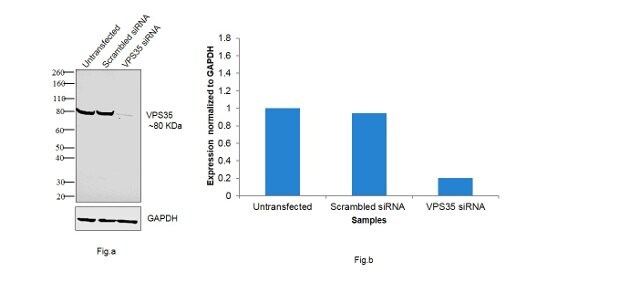
- KD of VPS35 was achieved by transfecting HeLa with VPS35 specific siRNAs (Silencer® select Product # s31375, s31376). Western blot analysis (Fig. a) was performed using whole cell extracts from the VPS35 KD cells (Lane 3), non-specific scrambled siRNA transfected cells (Lane 2) and untransfected cells (Lane 1). The blot was probed with VPS35 Polyclonal Antibody (Product # PA5-21898, 1:1000 dilution) and Goat anti-Rabbit IgG (H+L) Superclonal™ Secondary Antibody, HRP conjugate (Product # A27036, 1:4000 dilution). Densitometric analysis of this western blot is shown in histogram (Fig. b). Decrease in signal upon siRNA mediated knock down confirms that antibody is specific to VPS35. .
- Submitted by
- Invitrogen Antibodies (provider)
- Main image

- Experimental details
- Western blot was performed using Anti-VPS35 Rabbit Polyclonal Antibody (Product # PA5-21898) and 80 kDa band corresponding to VPS35 was observed across the cell lines tested. Whole cell extracts (30 µg lysate) of U-87 MG (Lane 1), A549 (Lane 2), U-937 (Lane 3), SH-SY5Y (Lane 4), HeLa (Lane 5), tissue extracts of Mouse Lung (Lane 6), Rat Lung (Lane 7) and Mouse Spleen (Lane 8) were electrophoresed using Novex® NuPAGE® 4-12% Bis-Tris gel (Product # NP0322BOX). Resolved proteins were then transferred onto a nitrocellulose membrane (Product # IB23001) by iBlot® 2 Dry Blotting System (Product # IB21001). The blot was probed with the primary antibody (1:1000 Dilution) and detected by chemiluminescence Goat Anti-Rabbit IgG Secondary Antibody, HRP conjugate (Product # A27036, 1:4000 dilution) using the iBright FL 1000 (Product # A32752). Chemiluminescent detection was performed using Novex® ECL Chemiluminescent Substrate Reagent Kit (Product # WP20005).

Supportive validation
- Submitted by
- Invitrogen Antibodies (provider)
- Main image
- Experimental details
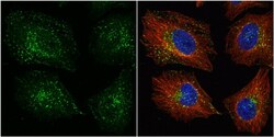
- Immunocytochemistry-Immunofluorescence analysis of VPS35 was performed in HeLa cells fixed in 4% paraformaldehyde at RT for 15 min. Green: VPS35 Polyclonal Antibody (Product # PA5-21898) diluted at 1:1000. Red: alpha Tubulin, a cytoskeleton marker. Blue: Hoechst 33342 staining.

- Submitted by
- Invitrogen Antibodies (provider)
- Main image
- Experimental details
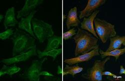
- VPS35 antibody detects VPS35 protein at endosome by immunofluorescent analysis. Sample: HeLa cells were fixed in 4% paraformaldehyde at RT for 15 min. Green: VPS35 stained by VPS35 antibody (Product # PA5-21898) diluted at 1:1,000. Red: alpha Tubulin, a cytoskeleton marker, stained by alpha Tubulin Polyclonal Antibody [GT114] (Product # MA5-31466) diluted at 1:1,000. Blue: Fluoroshield with DAPI .

- Submitted by
- Invitrogen Antibodies (provider)
- Main image
- Experimental details
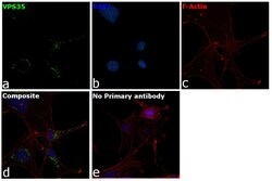
- Immunofluorescence analysis of VPS35 was performed using U-87 MG cells. The cells were fixed with 4% paraformaldehyde for 10 minutes, permeabilized with 0.1% Triton™ X-100 for 10 minutes, and blocked with 1% BSA for 1 hour at room temperature. The cells were labeled with VPS35 Rabbit Polyclonal Antibody (Product # PA5-21898) at 1:200 dilution in 0.1% BSA and incubated overnight at 4 degree and then labeled with Goat anti-Rabbit IgG (H+L) Superclonal™ Secondary Antibody, Alexa Fluor® 488 conjugate (Product # A27034) at a dilution of 1:2000 for 45 minutes at room temperature (Panel a: green). ). Nuclei (Panel b: blue) were stained with ProLong™ Diamond Antifade Mountant with DAPI (Product # P36962). F-actin (Panel c: red) was stained with Rhodamine Phalloidin (Product # R415, 1:300). Panel d represents the composite image showing endosomal localization. Panel e represents control cells with no primary antibody to assess background. The images were captured at 60X magnification. .

Supportive validation
- Submitted by
- Invitrogen Antibodies (provider)
- Main image

- Experimental details
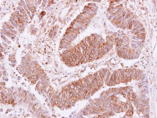
- VPS35 Polyclonal Antibody detects VPS35 protein at cytoplasm and membrane on human colon carcinoma by immunohistochemical analysis. Sample: Paraffin-embedded colon carcinoma. VPS35 Polyclonal Antibody (Product # PA5-21898) dilution: 1:250. Antigen Retrieval: EDTA based buffer, pH 8.0, 15 min.
Supportive validation
- Submitted by
- Invitrogen Antibodies (provider)
- Main image

- Experimental details
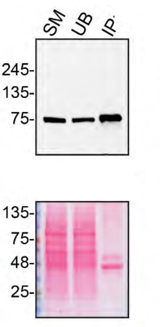
- Immunoprecipitation of VPS35 was performed on HAP1 WT cell lysate. Antibody-bead conjugate was prepared by adding 2 µg of VPS35 Polyclonal Antibody (Product # PA5-21898) with 30 µL of Dynabeads™ Protein A (Product # 10002D) and rocked for ~1 hour at 4 degree celcius. One mg of protein was incubated with the antibody-bead conjugate for ~2 hours at 4 degree celcius. Following centrifugation and multiple washes, 2% starting material (SM), 2% unbound fraction (UB) and immunoprecipitated fraction (IP) were processed for immunoblot using a different antibody. Ponceau stained transfer of blot is shown (below immunoblot). Data courtesy of YCharOS Inc., an open science company with the mission of characterizing commercially available antibodies using knockout validation.