 Explore
 Validate
 Learn
Learn Western blot
 Immunocytochemistry
Antibody data
- Antibody Data
- Antigen structure
- References [0]
- Comments [0]
- Validations
- Immunocytochemistry [3]
- Immunohistochemistry [1]
Submit
Validation data
Reference
Comment
Report error
- Product number
- SP5230P - Provider product page

- Provider
- Acris Antibodies GmbH
- Proper citation
- Acris Antibodies GmbH Cat#SP5230P, RRID:AB_980812
- Product name
- anti Collagen type XVIII alpha 1 chain
- Antibody type
- Polyclonal
- Antigen
- Synthetic Peptide: R(129) R L M/T E S Y C E T W R T E(142). SP5230P immunizing peptides are two peptides derived from Mouse and Human Endostatin protein corresponding to amino acid residues 129-142 of Endostatin.
- Reactivity
- Human, Mouse
- Host
- Rabbit
- Vial size
- 0.1 mg
- Concentration
- 1.0 mg/ml
No comments: Submit comment
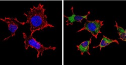
Supportive validation
- Submitted by
- Acris Antibodies GmbH (provider)
- Main image

- Experimental details
- Immunofluorescent analysis of Endostatin (green) showing positive staining in the secretion of A431 cells (right) compared with a negative control in the absence of primary antibody (left). Formalin-fixed cells were permeabilized with 0.1% Triton X-100 in TBS for 5-10 minutes, blocked with 3% BSA-PBS for 30 minutes at room temperature and probed with an Endostatin polyclonal antibody (Cat.-No SP5230P) in 3% BSA-PBS at a dilution of 1:200 and incubated overnight at 4 ºC in a humidified chamber. Cells were washed with PBST and incubated with a DyLight 488-conjugated goat-anti-rabbit IgG secondary antibody in PBS at room temperature in the dark. F-actin (red) was stained with a flourescent red phalloidin and nuclei (blue) were stained with DAPI for 5-10 minutes in the dark. Images were taken at a magnification of 60x.

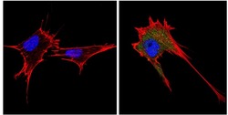
- Submitted by
- Acris Antibodies GmbH (provider)
- Main image
- Experimental details
- Immunofluorescent analysis of Endostatin (green) showing positive staining in the secretion of 293T cells (right) compared with a negative control in the absence of primary antibody (left). Formalin-fixed cells were permeabilized with 0.1% Triton X-100 in TBS for 5-10 minutes, blocked with 3% BSA-PBS for 30 minutes at room temperature and probed with an Endostatin polyclonal antibody (Cat.-No SP5230P) in 3% BSA-PBS at a dilution of 1:200 and incubated overnight at 4 ºC in a humidified chamber. Cells were washed with PBST and incubated with a DyLight 488-conjugated goat-anti-rabbit IgG secondary antibody in PBS at room temperature in the dark. F-actin (red) was stained with a flourescent red phalloidin and nuclei (blue) were stained with DAPI for 5-10 minutes in the dark. Images were taken at a magnification of 60x.

- Submitted by
- Acris Antibodies GmbH (provider)
- Main image
- Experimental details
- Immunofluorescent analysis of Endostatin (green) showing positive staining in the secretion of NIH-3T3 cells (right) compared with a negative control in the absence of primary antibody (left). Formalin-fixed cells were permeabilized with 0.1% Triton X-100 in TBS for 5-10 minutes, blocked with 3% BSA-PBS for 30 minutes at room temperature and probed with an Endostatin polyclonal antibody (Cat.-No SP5230P) in 3% BSA-PBS at a dilution of 1:200 and incubated overnight at 4 ºC in a humidified chamber. Cells were washed with PBST and incubated with a DyLight 488-conjugated goat-anti-rabbit IgG secondary antibody in PBS at room temperature in the dark. F-actin (red) was stained with a flourescent red phalloidin and nuclei (blue) were stained with DAPI for 5-10 minutes in the dark. Images were taken at a magnification of 60x.

Supportive validation
- Submitted by
- Acris Antibodies GmbH (provider)
- Main image
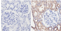
- Experimental details
- Immunohistochemistry analysis of Endostatin showing positive staining in the secretion of paraffin-treated Human kidney tissue (right) compared with a negative control in the absence of primary antibody (left). To expose target proteins, antigen retrieval method was performed using 10mM sodium citrate (pH 6.0), microwaved for 8-15 min. Following antigen retrieval, tissues were blocked in 3% H2O2-methanol for 15 min at room temperature, washed with ddH2O and PBS, and then probed with an endostatin polyclonal antibody (Cat.-No SP5230P) diluted by 3% BSA-PBS at a dilution of 1:100 overnight at 4°C in a humidified chamber. Tissues were washed extensively PBST and detection was performed using an HRP-conjugated secondary antibody followed by colorimetric detection using a DAB kit. Tissues were counterstained with hematoxylin and dehydrated with ethanol and xylene to prep for mounting.
