Explore
 Validate
 Learn
Learn Western blot
 ELISA
ELISAAntibody data
- Antibody Data
- Antigen structure
- References [0]
- Comments [0]
- Validations
- Western blot [4]
- Immunocytochemistry [2]
- Immunohistochemistry [2]
Submit
Validation data
Reference
Comment
Report error
- Product number
- MA5-33221 - Provider product page

- Provider
- Invitrogen Antibodies
- Product name
- FTO Recombinant Rabbit Monoclonal Antibody (4G9)
- Antibody type
- Monoclonal
- Antigen
- Synthetic peptide
- Reactivity
- Human
- Host
- Rabbit
- Isotype
- IgG
- Antibody clone number
- 4G9
- Vial size
- 100 µL
- Concentration
- 0.26 mg/mL
- Storage
- -20°C or -80°C if preferred
No comments: Submit comment
Supportive validation
- Submitted by
- Invitrogen Antibodies (provider)
- Main image

- Experimental details
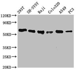
- Western Blot analysis of FTO using a FTO Monoclonal antibody (Product # MA5-33221) at a concentration of 0.7 µg/mL. Positive WB detected in: 293T whole cell lysate, SH-SY5Y whole cell lysate, Raji whole cell lysate, Colo320 whole cell lysate, A549 whole cell lysate, PC3 whole cell lysate. A secondary Goat polyclonal antibody to rabbit IgG was applied at a 1:50,000 dilution. Observed band size: 59 kDa.

- Submitted by
- Invitrogen Antibodies (provider)
- Main image
- Experimental details
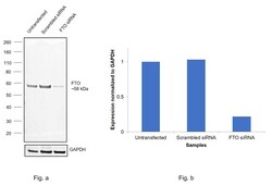
- Knockdown of Alpha-ketoglutarate-dependent dioxygenase FTO was achieved by transfecting SH-SY5Y with Alpha-ketoglutarate-dependent dioxygenase FTO specific siRNAs (Silencer® select Product # S35512, S35511). Western blot analysis (Fig. a) was performed using Nuclear enriched extracts from the Alpha-ketoglutarate-dependent dioxygenase FTO knockdown cells (lane 3), non-targeting scrambled siRNA transfected cells (lane 2) and untransfected cells (lane 1). The blot was probed with FTO Recombinant Rabbit Monoclonal Antibody (Product # MA5-33221, 1:3000) and Goat anti-Rabbit IgG (H+L) Superclonal™ Recombinant Secondary Antibody, HRP (Product # A27036, 1:4000). Densitometric analysis of this western blot is shown in histogram (Fig. b). Decrease in signal upon siRNA mediated knock down confirms that antibody is specific to Alpha-ketoglutarate-dependent dioxygenase FTO.

- Submitted by
- Invitrogen Antibodies (provider)
- Main image
- Experimental details
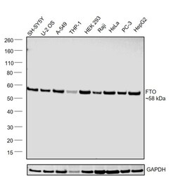
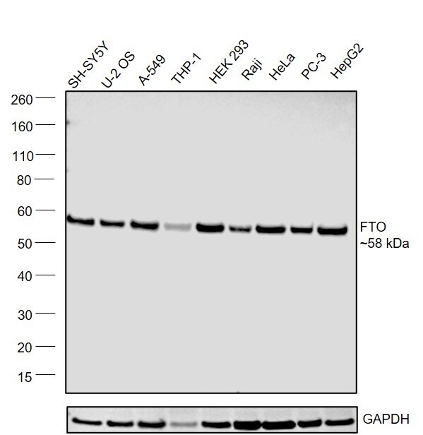
- Western blot was performed using Anti-FTO Recombinant Rabbit Monoclonal Antibody (Product # MA5-33221) and a ~58kDa band corresponding to Alpha-ketoglutarate-dependent dioxygenase FTO was observed across cell lines tested . Nuclear enriched extracts (30 µg lysate) of SH-SY5Y (Lane 1), U-2 OS (Lane 2), A549 (Lane 3), THP-1 (Lane 4), HEK-293 (Lane 5), Raji (Lane 6), HeLa (Lane 7), PC-3 (Lane 8), Hep G2 (Lane 9) were electrophoresed using NuPAGE™ 4-12% Bis-Tris Protein Gel (Product # NP0321BOX). Resolved proteins were then transferred onto a Nitrocellulose membrane (Product # LC2001) by iBlot® 2 Dry Blotting System (Product # IB21001). The blot was probed with the primary antibody (1:2500) and detected by chemiluminescence with Goat anti-Rabbit IgG (H+L) Superclonal™ Recombinant Secondary Antibody, HRP (Product # A27036, 1:4000) using the iBright FL 1000 (Product # A32752). Chemiluminescent detection was performed using Novex® ECL Chemiluminescent Substrate Reagent Kit (Product # WP20005).
Supportive validation
- Submitted by
- Invitrogen Antibodies (provider)
- Main image
- Experimental details
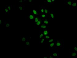
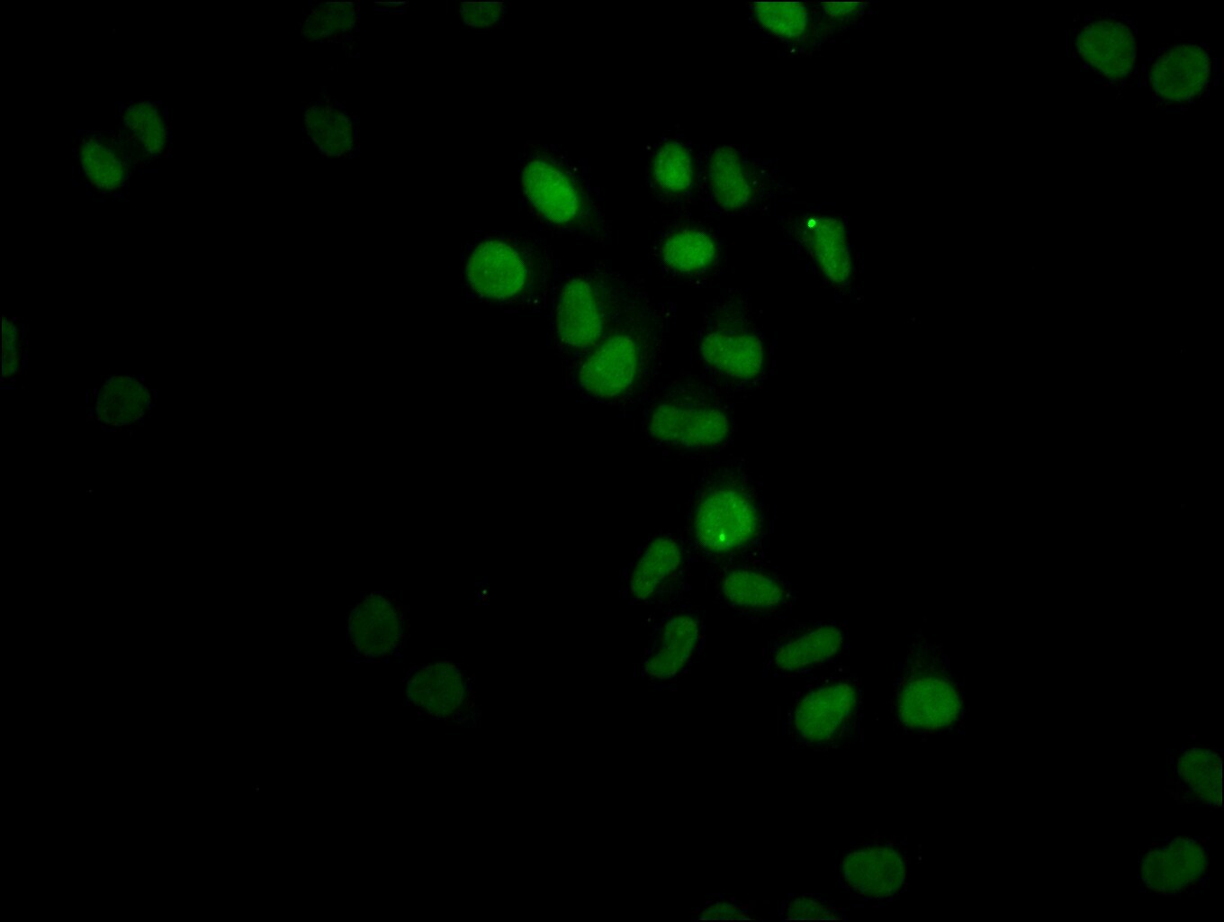
- Immunofluorescent analysis of FTO in Hela cells using a FTO monoclonal antibody (Product # MA5-33221) at a dilution of 1:23. The cells were fixed in 4% formaldehyde, permeabilized using 0.2% Triton X-100 and blocked in 10% normal Goat Serum. The cells were then incubated with the antibody overnight at 4°C. The secondary antibody was Alexa Fluor 488-congugated Goat Anti-Rabbit IgG (H+L). Cells were counter-stained with DAPI.
- Submitted by
- Invitrogen Antibodies (provider)
- Main image
- Experimental details
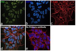
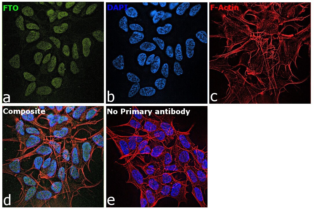
- Immunofluorescence analysis of Alpha-ketoglutarate-dependent dioxygenase FTO was performed using 70% confluent log phase SH-SY5Y cells. The cells were fixed with 4% paraformaldehyde for 10 minutes, permeabilized with 0.1% Triton™ X-100 for 15 minutes, and blocked with 2% BSA for 45 minutes at room temperature. The cells were labeled with FTO Recombinant Rabbit Monoclonal Antibody (Product # MA5-33221) at 1:100 in 0.1% BSA, incubated at 4 degree celsius overnight and then labeled with Donkey anti-Rabbit IgG (H+L) Highly Cross-Adsorbed Secondary Antibody, Alexa Fluor Plus 488 (Product # A32790), (1:2000), for 45 minutes at room temperature (Panel a: Green). Nuclei (Panel b:Blue) were stained with ProLong™ Diamond Antifade Mountant with DAPI (Product # P36962). F-actin (Panel c: Red) was stained with Rhodamine Phalloidin (Product # R415, 1:300). Panel d represents the merged image showing Nucleus and cytoplasm localization. Panel e represents control cells with no primary antibody to assess background. The images were captured at 60X magnification.
Supportive validation
- Submitted by
- Invitrogen Antibodies (provider)
- Main image
- Experimental details
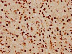
- Immunohistochemical analysis of FTO in paraffin embedded human glioma cancer using a FTO monoclonal antibody (Product # MA5-33221) at a dilution of 1:70. After dewaxing and hydration, antigen retrieval was mediated by high pressure in a citrate buffer (pH 6.0). Section was blocked with 10% normal goat serum 30min at RT. Then primary antibody (1% BSA) was incubated at 4°C overnight. The primary is detected by a biotinylated secondary antibody and visualized using an HRP conjugated SP system.

- Submitted by
- Invitrogen Antibodies (provider)
- Main image

- Experimental details
- Immunohistochemical analysis of FTO in paraffin embedded human skeletal muscle tissue using a FTO monoclonal antibody (Product # MA5-33221) at a dilution of 1:70. After dewaxing and hydration, antigen retrieval was mediated by high pressure in a citrate buffer (pH 6.0). Section was blocked with 10% normal goat serum 30min at RT. Then primary antibody (1% BSA) was incubated at 4°C overnight. The primary is detected by a biotinylated secondary antibody and visualized using an HRP conjugated SP system.