 Explore
 Validate
 Learn
Learn Western blot
 Immunocytochemistry
Antibody data
- Antibody Data
- Antigen structure
- References [1]
- Comments [0]
- Validations
- Western blot [1]
- Immunocytochemistry [1]
- Immunohistochemistry [9]
Submit
Validation data
Reference
Comment
Report error
- Product number
- HPA041863 - Provider product page

- Provider
- Atlas Antibodies
- Proper citation
- Atlas Antibodies Cat#HPA041863, RRID:AB_10794638
- Product name
- Anti-UQCRFS1
- Antibody type
- Polyclonal
- Reactivity
- Human, Rat
- Host
- Rabbit
- Conjugate
- Unconjugated
- Antigen sequence
RVKKPEWVILIGVCTHLGCVPIANAGDFGGYYCPC
HGSHYDASGRIRLGPAPLNLEVPTYEFTSDDMVIV- Isotype
- IgG
- Vial size
- 100 µl
- Storage
- Store at +4°C for short term storage. Long time storage is recommended at -20°C.
Submitted references C11orf83, a mitochondrial cardiolipin-binding protein involved in bc1 complex assembly and supercomplex stabilization.
Desmurs M, Foti M, Raemy E, Vaz FM, Martinou JC, Bairoch A, Lane L
Molecular and cellular biology 2015 Apr;35(7):1139-56
Molecular and cellular biology 2015 Apr;35(7):1139-56
No comments: Submit comment
Enhanced validation
- Submitted by
- Atlas Antibodies (provider)
- Enhanced method
- Independent antibody validation
- Main image

- Experimental details
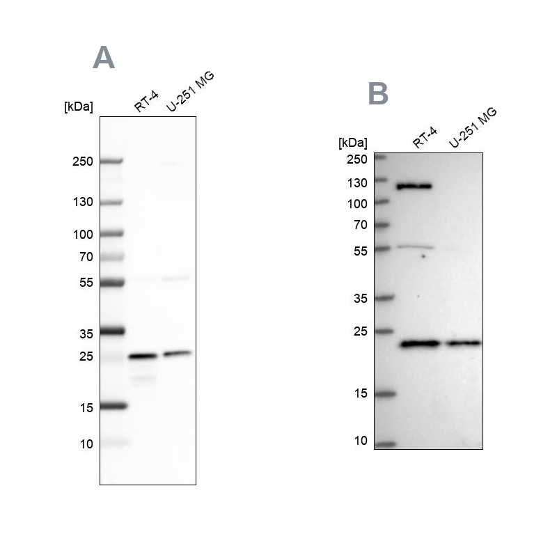
- Western blot analysis using Anti-UQCRFS1 antibody HPA041863 (A) shows similar pattern to independent antibody HPA050339 (B).
Supportive validation
- Submitted by
- Atlas Antibodies (provider)
- Main image

- Experimental details
- Immunofluorescent staining of human cell line U-251 MG shows localization to mitochondria.
- Sample type
- HUMAN

Enhanced validation
Enhanced validation
Supportive validation
- Submitted by
- Atlas Antibodies (provider)
- Enhanced method
- Orthogonal validation
- Main image
- Experimental details
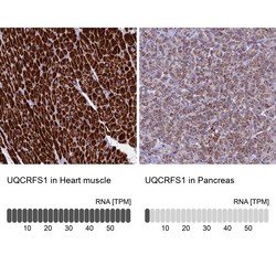
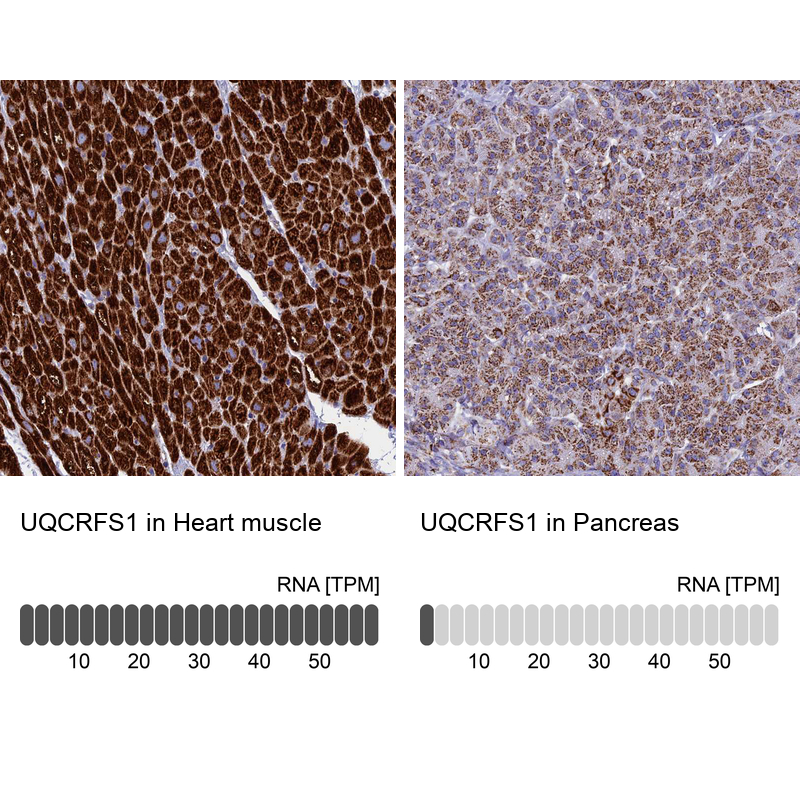
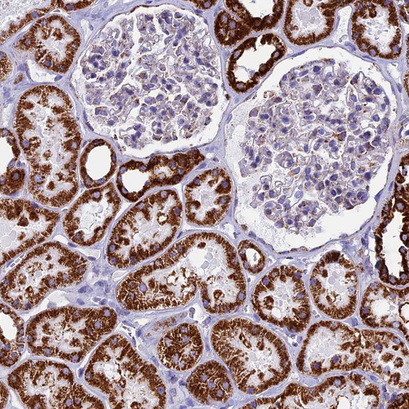
- Immunohistochemistry analysis in human heart muscle and pancreas tissues using HPA041863 antibody. Corresponding UQCRFS1 RNA-seq data are presented for the same tissues.
- Sample type
- HUMAN
Enhanced validation
- Submitted by
- Atlas Antibodies (provider)
- Enhanced method
- Independent antibody validation
- Main image

- Experimental details
- Immunohistochemical staining of human colon, kidney, liver and testis using Anti-UQCRFS1 antibody HPA041863 (A) shows similar protein distribution across tissues to independent antibody HPA050339 (B).

Supportive validation
- Submitted by
- Atlas Antibodies (provider)
- Main image
- Experimental details
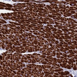
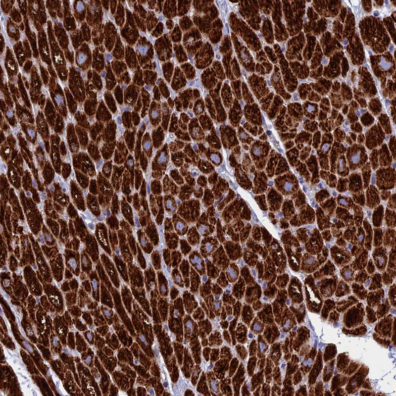
- Immunohistochemical staining of human heart muscle shows strong cytoplasmic positivity in myocytes.

- Submitted by
- Atlas Antibodies (provider)
- Main image

- Experimental details
- Immunohistochemical staining of human heart muscle shows moderate to strong cytoplasmic positivity in myocytes.
- Sample type
- HUMAN
- Submitted by
- Atlas Antibodies (provider)
- Main image
- Experimental details
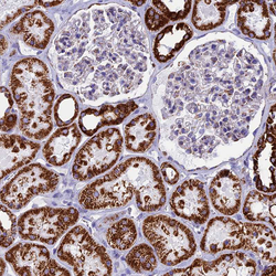
- Immunohistochemical staining of human kidney shows moderate to strong cytoplasmic positivity in cells in tubules.
- Sample type
- HUMAN
- Submitted by
- Atlas Antibodies (provider)
- Main image

- Experimental details
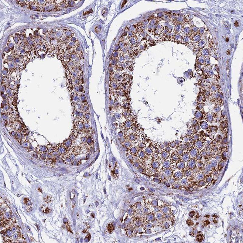
- Immunohistochemical staining of human testis shows moderate to strong cytoplasmic positivity in cells in seminiferous ducts and in Leydig cells.
- Sample type
- HUMAN
- Submitted by
- Atlas Antibodies (provider)
- Main image
- Experimental details
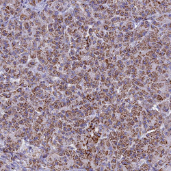
- Immunohistochemical staining of human pancreas shows moderate to strong cytoplasmic positivity in exocrine glandular cells.
- Sample type
- HUMAN

- Submitted by
- Atlas Antibodies (provider)
- Main image
- Experimental details
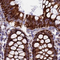
- Immunohistochemical staining of human colon using Anti-UQCRFS1 antibody HPA041863.
- Sample type
- HUMAN

- Submitted by
- Atlas Antibodies (provider)
- Main image

- Experimental details
- Immunohistochemical staining of human liver using Anti-UQCRFS1 antibody HPA041863.
- Sample type
- HUMAN
