Explore
 Validate
 Learn
Learn Western blot
 Immunoprecipitation
Antibody data
- Antibody Data
- Antigen structure
- References [1]
- Comments [0]
- Validations
- Western blot [6]
- Immunocytochemistry [4]
- Immunohistochemistry [5]
- Other assay [1]
Submit
Validation data
Reference
Comment
Report error
- Product number
- PA5-34911 - Provider product page

- Provider
- Invitrogen Antibodies
- Product name
- USP7 Polyclonal Antibody
- Antibody type
- Polyclonal
- Antigen
- Recombinant protein fragment
- Description
- Recommended positive controls: 293T, A431, HeLa, HepG2, USP7 transfected 293T lysate, NIH-3T3, JC, PC-12. Predicted reactivity: Mouse (97%), Rat (97%), Xenopus laevis (93%), Pig (97%), Chicken (93%). Store product as a concentrated solution. Centrifuge briefly prior to opening the vial.
- Reactivity
- Human, Mouse, Rat
- Host
- Rabbit
- Isotype
- IgG
- Vial size
- 100 µL
- Concentration
- 0.94 mg/mL
- Storage
- Store at 4°C short term. For long term storage, store at -20°C, avoiding freeze/thaw cycles.
Submitted references Reversal of viral and epigenetic HLA class I repression in Merkel cell carcinoma.
Lee PC, Klaeger S, Le PM, Korthauer K, Cheng J, Ananthapadmanabhan V, Frost TC, Stevens JD, Wong AY, Iorgulescu JB, Tarren AY, Chea VA, Carulli IP, Lemvigh CK, Pedersen CB, Gartin AK, Sarkizova S, Wright KT, Li LW, Nomburg J, Li S, Huang T, Liu X, Pomerance L, Doherty LM, Apffel AM, Wallace LJ, Rachimi S, Felt KD, Wolff JO, Witten E, Zhang W, Neuberg D, Lane WJ, Zhang G, Olsen LR, Thakuria M, Rodig SJ, Clauser KR, Starrett GJ, Doench JG, Buhrlage SJ, Carr SA, DeCaprio JA, Wu CJ, Keskin DB
The Journal of clinical investigation 2022 Jul 1;132(13)
The Journal of clinical investigation 2022 Jul 1;132(13)
No comments: Submit comment
Supportive validation
- Submitted by
- Invitrogen Antibodies (provider)
- Main image
- Experimental details
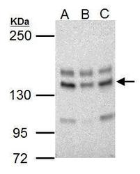
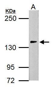
- Western Blot using USP7 Polyclonal Antibody (Product # PA5-34911). Sample (30 µg of whole cell lysate). Lane A: NIH-3T3. Lane B: JC. Lane C: BCL-1. 5% SDS PAGE. USP7 Polyclonal Antibody (Product # PA5-34911) diluted at 1:5,000. The HRP-conjugated anti-rabbit IgG antibody was used to detect the primary antibody.
- Submitted by
- Invitrogen Antibodies (provider)
- Main image
- Experimental details
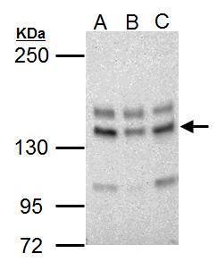
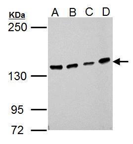
- Western Blot using USP7 Polyclonal Antibody (Product # PA5-34911). Sample (30 µg of whole cell lysate). Lane A: 293T. Lane B: A431. Lane C: HeLa. Lane D: HepG2. 5% SDS PAGE. USP7 Polyclonal Antibody (Product # PA5-34911) diluted at 1:1,000. The HRP-conjugated anti-rabbit IgG antibody was used to detect the primary antibody.
- Submitted by
- Invitrogen Antibodies (provider)
- Main image
- Experimental details
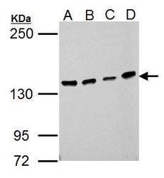
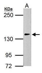
- Western Blot using USP7 Polyclonal Antibody (Product # PA5-34911). Sample (30 µg of whole cell lysate). Lane A: PC-12. 5% SDS PAGE. USP7 Polyclonal Antibody (Product # PA5-34911) diluted at 1:5,000. The HRP-conjugated anti-rabbit IgG antibody was used to detect the primary antibody.
- Submitted by
- Invitrogen Antibodies (provider)
- Main image
- Experimental details
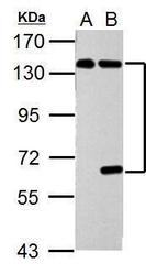
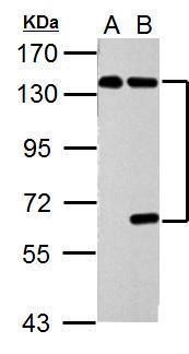
- Western Blot using USP7 Polyclonal Antibody (Product # PA5-34911). Sample (30 µg of whole cell lysate). Lane A: Non-transfected 293T lysates. Lane B: USP7 transfected 293T lysates. 7.5% SDS PAGE. USP7 Polyclonal Antibody (Product # PA5-34911) diluted at 1:5,000. The HRP-conjugated anti-rabbit IgG antibody was used to detect the primary antibody.
- Submitted by
- Invitrogen Antibodies (provider)
- Main image

- Experimental details
- Knockdown of USP7 was achieved by transfecting A549 with USP7 specific siRNAs (Silencer® select Product # s15440). Western blot analysis (Fig. a) was performed using whole cell extracts from the USP7 knockdown cells (lane 3), non-specific scrambled siRNA transfected cells (lane 2) and untransfected cells (lane 1). The blots were probed with USP7 Polyclonal Antibody (Product # PA5-34911, 1:1000 dilution) and Goat anti-Rabbit IgG (H+L) Superclonal™ Secondary Antibody, HRP conjugate (Product # A27036, 0.25µg/ml, 1:4000 dilution). Densitometric analysis of this western blot is shown in histogram (Fig. b). Decrease in signal upon siRNA mediated knock down confirms that antibody is specific to USP7.

- Submitted by
- Invitrogen Antibodies (provider)
- Main image
- Experimental details
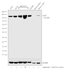
- Western blot analysis was performed on whole cell extracts (30 µg lysate) of A549 (Lane 1), HT29 (Lane 2), HCT116 (Lane 3), HCT116 treated with Metformin (5mM for 12 hours) (Lane 4), SW480 (Lane 5), tissue extracts of Mouse spleen (Lane 6) and Mouse colon (Lane 7). The blot was probed with Anti-USP7 Polyclonal Antibody (Product # PA5-34911, 1:1000 dilution) and detected by chemiluminescence using Goat anti-Rabbit IgG (H+L) Superclonal™ Secondary Antibody, HRP conjugate (Product # A27036, 0.25 µg/ml, 1:4000 dilution). A 128 kDa band corresponding to USP7 was observed across all the cell lines and tissue positive for USP7 and overexpressed in HCT116 cells upon treatment with Metformin. This band was absent in Mouse spleen which is reported to be negative.

Supportive validation
- Submitted by
- Invitrogen Antibodies (provider)
- Main image
- Experimental details
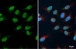
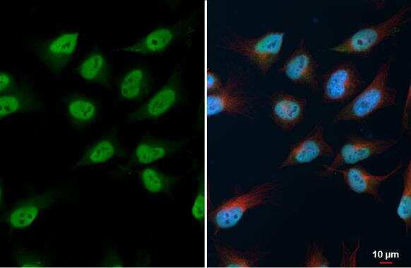
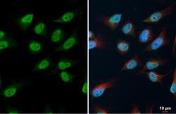
- USP7 Polyclonal Antibody detects USP7 protein at nucleus by immunofluorescent analysis. Sample: HeLa cells were fixed in 4% paraformaldehyde at RT for 15 min. Green: USP7 stained by USP7 Polyclonal Antibody (Product # PA5-34911) diluted at 1:500. Red: alpha Tubulin, a cytoskeleton marker, stained by alpha Tubulin antibody [GT114] (Product # MA5-31466) diluted at 1:1,000. Blue: Fluoroshield with DAPI .
- Submitted by
- Invitrogen Antibodies (provider)
- Main image
- Experimental details
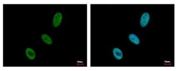
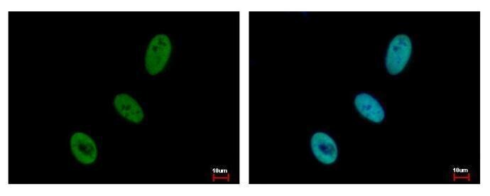
- USP7 Polyclonal Antibody detects USP7 protein at nucleus by immunofluorescent analysis. Sample: HeLa cells were fixed in 4% paraformaldehyde at RT for 15 min. Green: USP7 protein stained by USP7 Polyclonal Antibody (Product # PA5-34911) diluted at 1:500. Blue: Hoechst 33343 staining.
- Submitted by
- Invitrogen Antibodies (provider)
- Main image
- Experimental details
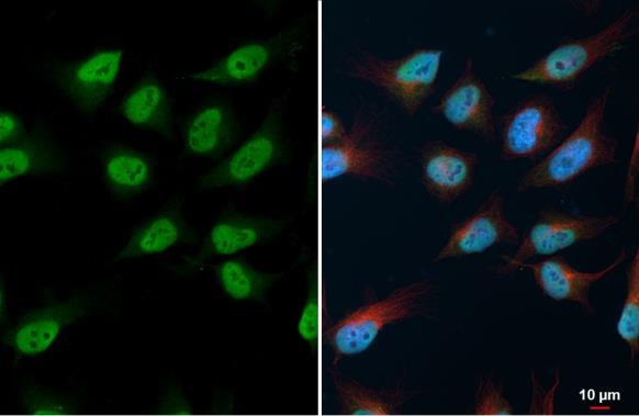
- USP7 Polyclonal Antibody detects USP7 protein at nucleus by immunofluorescent analysis. Sample: HeLa cells were fixed in 4% paraformaldehyde at RT for 15 min. Green: USP7 stained by USP7 Polyclonal Antibody (Product # PA5-34911) diluted at 1:500. Red: alpha Tubulin, a cytoskeleton marker, stained by alpha Tubulin antibody [GT114] (Product # MA5-31466) diluted at 1:1,000. Blue: Fluoroshield with DAPI .
- Submitted by
- Invitrogen Antibodies (provider)
- Main image
- Experimental details
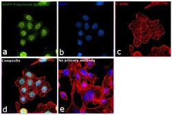
- Immunofluorescence analysis of USP7 was performed using 70% confluent log phase SW480 cells. The cells were fixed with 4% paraformaldehyde for 10 minutes, permeabilized with 0.1% Triton™ X-100 for 15 minutes, and blocked with 1% BSA for 1 hour at room temperature. The cells were labeled with USP7 Rabbit Polyclonal Antibody (Product # PA5-34911) at 1:200 dilution in 0.1% BSA, incubated at 4 degree Celsius overnight and then labeled with Goat anti-Rabbit IgG (H+L) Superclonal™ Secondary Antibody, Alexa Fluor® 488 conjugate (Product # A27034) at a dilution of 1:2000 for 45 minutes at room temperature (Panel a: green). Nuclei (Panel b: blue) were stained with ProLong™ Diamond Antifade Mountant with DAPI (Product # P36962). F-actin (Panel c: red) was stained with Rhodamine Phalloidin (Product # R415, 1:300). Panel d represents the merged image showing predominantly nuclear localization. Panel e represents control cells with no primary antibody to assess background. The images were captured at 60X magnification.
Supportive validation
- Submitted by
- Invitrogen Antibodies (provider)
- Main image
- Experimental details
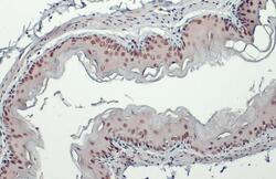
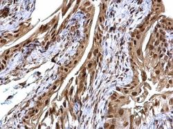
- USP7 Polyclonal Antibody detects USP7 protein at cytoplasm and nucleus by immunohistochemical analysis. Sample: Paraffin-embedded mouse esophagus. USP7 stained by USP7 Polyclonal Antibody (Product # PA5-34911) diluted at 1:500. Antigen Retrieval: Citrate buffer, pH 6.0, 15 min.

- Submitted by
- Invitrogen Antibodies (provider)
- Main image
- Experimental details
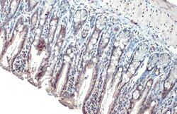
- USP7 Polyclonal Antibody detects USP7 protein at cytoplasm and nucleus by immunohistochemical analysis. Sample: Paraffin-embedded mouse intestine. USP7 stained by USP7 Polyclonal Antibody (Product # PA5-34911) diluted at 1:500. Antigen Retrieval: Citrate buffer, pH 6.0, 15 min.

- Submitted by
- Invitrogen Antibodies (provider)
- Main image
- Experimental details
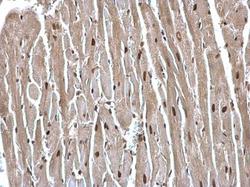
- USP7 Polyclonal Antibody detects USP7 protein at cytosol and nucleus on mouse heart by immunohistochemical analysis. Sample: Paraffin-embedded mouse heart. USP7 Polyclonal Antibody (Product # PA5-34911) dilution: 1:500. Antigen Retrieval: EDTA based buffer, pH 8.0, 15 min.
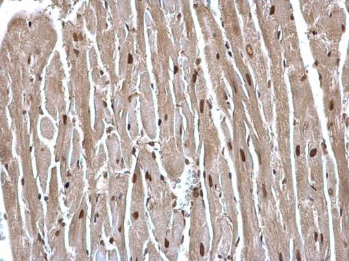
- Submitted by
- Invitrogen Antibodies (provider)
- Main image
- Experimental details
- USP7 Polyclonal Antibody detects USP7 protein at cytosol and nucleus on mouse heart by immunohistochemical analysis. Sample: Paraffin-embedded mouse heart. USP7 Polyclonal Antibody (Product # PA5-34911) dilution: 1:500. Antigen Retrieval: EDTA based buffer, pH 8.0, 15 min.

- Submitted by
- Invitrogen Antibodies (provider)
- Main image
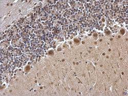
- Experimental details
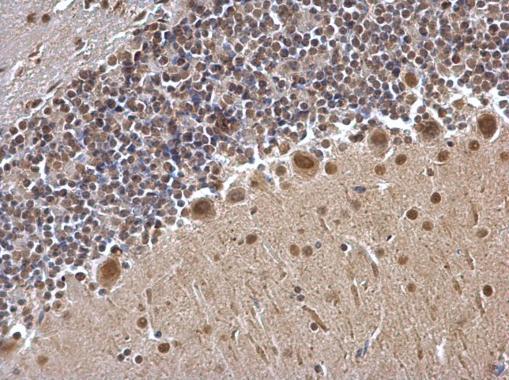
- USP7 Polyclonal Antibody detects USP7 protein at cytosol and nucleus on mouse hind brain by immunohistochemical analysis. Sample: Paraffin-embedded mouse hind brain. USP7 Polyclonal Antibody (Product # PA5-34911) dilution: 1:500. Antigen Retrieval: EDTA based buffer, pH 8.0, 15 min.
Supportive validation
- Submitted by
- Invitrogen Antibodies (provider)
- Main image
- Experimental details
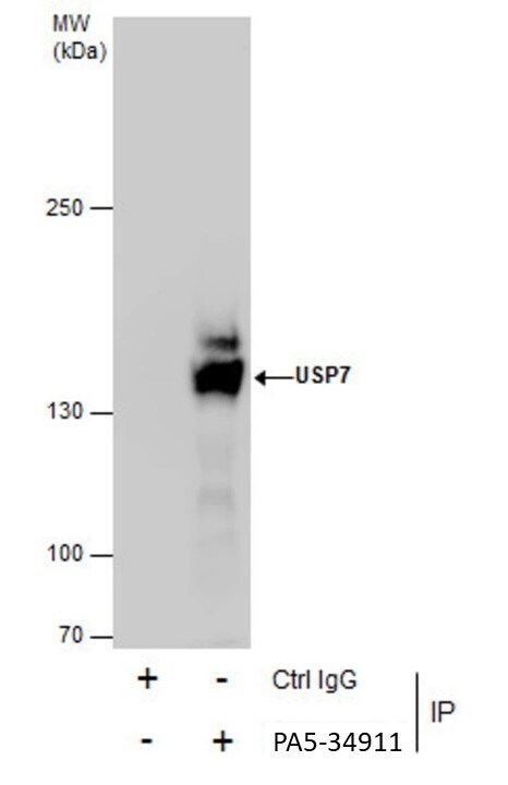
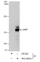
- Immunoprecipitation of USP7 was performed in HepG2 whole cell extracts using 5 µg of USP7 Polyclonal Antibody (Product # PA5-34911). Samples were transferred to a membrane and probed with USP7 Polyclonal Antibody as a primary antibody and an HRP-conjugated anti-Rabbit IgG was used as a secondary antibody.