 Explore
 Validate
 Learn
Learn Western blot
Antibody data
- Antibody Data
- Antigen structure
- References [2]
- Comments [0]
- Validations
- Western blot [4]
- Immunocytochemistry [1]
- Immunoprecipitation [1]
- Immunohistochemistry [3]
Submit
Validation data
Reference
Comment
Report error
- Product number
- GTX125894 - Provider product page

- Provider
- GeneTex
- Product name
- USP7 antibody
- Antibody type
- Polyclonal
- Reactivity
- Human, Mouse, Rat
- Host
- Rabbit
Submitted references Targeting USP7 Identifies a Metastasis-Competent State within Bone Marrow-Resident Melanoma CTCs.
Histone H2B monoubiquitination is a critical epigenetic switch for the regulation of autophagy.
Vishnoi M, Boral D, Liu H, Sprouse ML, Yin W, Goswami-Sewell D, Tetzlaff MT, Davies MA, Oliva ICG, Marchetti D
Cancer research 2018 Sep 15;78(18):5349-5362
Cancer research 2018 Sep 15;78(18):5349-5362
Histone H2B monoubiquitination is a critical epigenetic switch for the regulation of autophagy.
Chen S, Jing Y, Kang X, Yang L, Wang DL, Zhang W, Zhang L, Chen P, Chang JF, Yang XM, Sun FL
Nucleic acids research 2017 Feb 17;45(3):1144-1158
Nucleic acids research 2017 Feb 17;45(3):1144-1158
No comments: Submit comment
Supportive validation
- Submitted by
- GeneTex (provider)
- Main image
- Experimental details
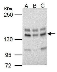
- Sample (30 ?g of whole cell lysate) A: NIH-3T3 B: JC C: BCL-1 5% SDS PAGE GTX125894 diluted at 1:5000 The HRP-conjugated anti-rabbit IgG antibody (GTX213110-01) was used to detect the primary antibody.
- Submitted by
- GeneTex (provider)
- Main image
- Experimental details
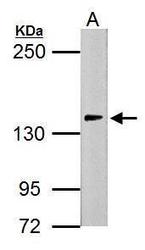
- Sample (30 ?g of whole cell lysate) A: PC-12 5% SDS PAGE GTX125894 diluted at 1:5000 The HRP-conjugated anti-rabbit IgG antibody (GTX213110-01) was used to detect the primary antibody.
- Submitted by
- GeneTex (provider)
- Main image
- Experimental details
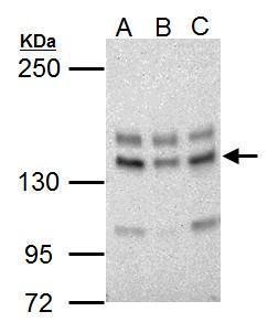
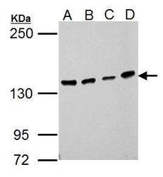
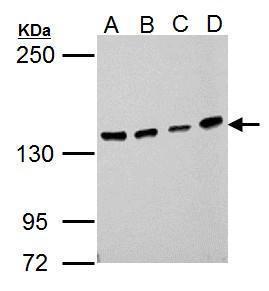
- Sample (30 ?g of whole cell lysate) A: 293T B: A431 C: HeLa D: HepG2 5% SDS PAGE GTX125894 diluted at 1:1000 The HRP-conjugated anti-rabbit IgG antibody (GTX213110-01) was used to detect the primary antibody.
- Submitted by
- GeneTex (provider)
- Main image
- Experimental details
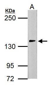
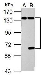
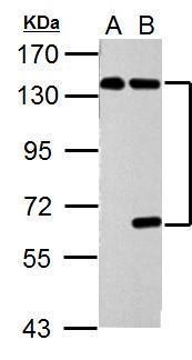
- Sample (30 ?g of whole cell lysate) A: ?Non-transfected 293T lysates? B: ?USP7 transfected 293T lysates 7.5% SDS PAGE GTX125894 diluted at 1:5000 The HRP-conjugated anti-rabbit IgG antibody (GTX213110-01) was used to detect the primary antibody.
Supportive validation
- Submitted by
- GeneTex (provider)
- Main image

- Experimental details
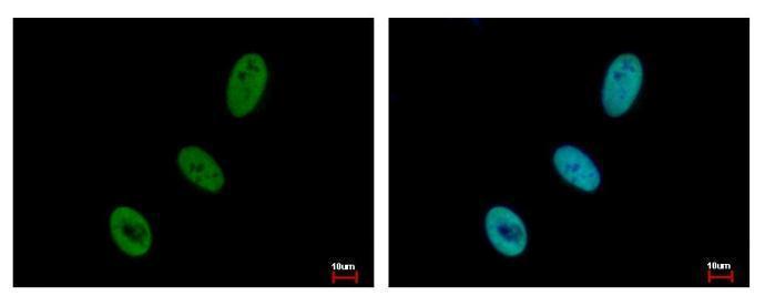
- USP7 antibody detects USP7 protein at nucleus by immunofluorescent analysis. Sample: HeLa cells were fixed in 4% paraformaldehyde at RT for 15 min.Green: USP7 protein stained by USP7 antibody (GTX125894) diluted at 1:500.Blue: Hoechst 33343 staining.
Supportive validation
- Submitted by
- GeneTex (provider)
- Main image
- Experimental details
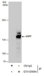
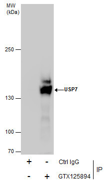
- Immunoprecipitation of USP7 protein from HepG2 whole cell extracts using 5 £gg of USP7 antibody (GTX125894).Western blot analysis was performed using USP7 antibody (GTX125894).EasyBlot anti-Rabbit IgG (GTX221666-01) was used as a secondary reagent.
Supportive validation
- Submitted by
- GeneTex (provider)
- Main image
- Experimental details
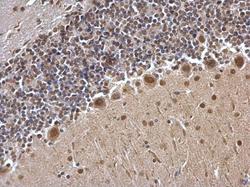
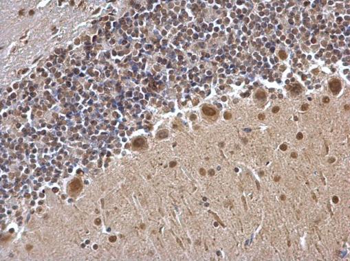
- USP7 antibody detects USP7 protein at cytosol and nucleus on mouse hind brain by immunohistochemical analysis. Sample: Paraffin-embedded mouse hind brain. USP7 antibody (GTX125894) dilution: 1:500.
- Submitted by
- GeneTex (provider)
- Main image
- Experimental details
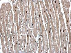
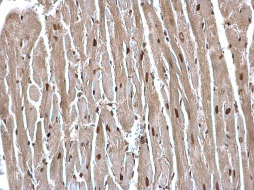
- USP7 antibody detects USP7 protein at cytosol and nucleus on mouse heart by immunohistochemical analysis. Sample: Paraffin-embedded mouse heart. USP7 antibody (GTX125894) dilution: 1:500.
- Submitted by
- GeneTex (provider)
- Main image
- Experimental details
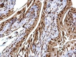
- USP7 antibody detects USP7 protein at cytosol and nucleus on mouse urinary bladder by immunohistochemical analysis. Sample: Paraffin-embedded mouse urinary bladder. USP7 antibody (GTX125894) dilution: 1:500.
