 Explore
 Validate
 Learn
Learn Western blot
 Immunohistochemistry
Antibody data
- Antibody Data
- Antigen structure
- References [2]
- Comments [0]
- Validations
- Immunohistochemistry [1]
Submit
Validation data
Reference
Comment
Report error
- Product number
- MAB134-050 - Provider product page

- Provider
- R&D Systems
- Product name
- Human WIF-1 Antibody
- Antibody type
- Monoclonal
- Description
- Protein A or G purified from hybridoma culture supernatant. Detects human WIF-1 in Western blots. In Western blots, less than 5% cross-reactivity with recombinant mouse WIF-1 is observed.
- Reactivity
- Human
- Host
- Mouse
- Conjugate
- Unconjugated
- Antigen sequence
AAD25402- Isotype
- IgG
- Antibody clone number
- 133015
- Vial size
- 50 ug
- Concentration
- LYOPH
- Storage
- Use a manual defrost freezer and avoid repeated freeze-thaw cycles. 12 months from date of receipt, -20 to -70 °C as supplied. 1 month, 2 to 8 °C under sterile conditions after reconstitution. 6 months, -20 to -70 °C under sterile conditions after reconstitution.
Submitted references Evidence for altered Wnt signaling in psoriatic skin.
Wnt pathway inhibitors are strongly down-regulated in pituitary tumors.
Gudjonsson JE, Johnston A, Stoll SW, Riblett MB, Xing X, Kochkodan JJ, Ding J, Nair RP, Aphale A, Voorhees JJ, Elder JT
The Journal of investigative dermatology 2010 Jul;130(7):1849-59
The Journal of investigative dermatology 2010 Jul;130(7):1849-59
Wnt pathway inhibitors are strongly down-regulated in pituitary tumors.
Elston MS, Gill AJ, Conaglen JV, Clarkson A, Shaw JM, Law AJ, Cook RJ, Little NS, Clifton-Bligh RJ, Robinson BG, McDonald KL
Endocrinology 2008 Mar;149(3):1235-42
Endocrinology 2008 Mar;149(3):1235-42
No comments: Submit comment
Supportive validation
- Submitted by
- R&D Systems (provider)
- Main image
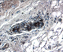
- Experimental details
- WIF-1 in Human Breast. WIF-1 was detected in immersion fixed paraffin-embedded sections of human breast using 8 µg/mL Mouse Anti-Human WIF-1 Monoclonal Antibody (Catalog # MAB134) overnight at 4 °C. Tissue was stained with the Anti-Mouse HRP-DAB Cell & Tissue Staining Kit (brown; Catalog # CTS002) and counterstained with hematoxylin (blue). View our protocol for Chromogenic IHC Staining of Paraffin-embedded Tissue Sections.
