 Explore
 Validate
 Learn
Learn Western blot
Antibody data
- Antibody Data
- Antigen structure
- References [0]
- Comments [0]
- Validations
- Western blot [1]
- Immunocytochemistry [4]
- Immunohistochemistry [10]
Submit
Validation data
Reference
Comment
Report error
- Product number
- GTX105504 - Provider product page

- Provider
- GeneTex
- Proper citation
- GeneTex Cat#GTX105504, RRID:AB_1950971
- Product name
- NAGLU antibody
- Antibody type
- Polyclonal
- Reactivity
- Human, Mouse, Rat
- Host
- Rabbit
No comments: Submit comment
Supportive validation
- Submitted by
- GeneTex (provider)
- Main image

- Experimental details
- Sample(30 ug whole cell lysate)A:H1299B:HeLa S3(GTX14654)C:Hep G2 (GTX27900)7.5% SDS PAGEGTX105504 diluted at 1:1000
- Validation comment
- WB

Supportive validation
- Submitted by
- GeneTex (provider)
- Main image

- Experimental details
- NAGLU antibody detects NAGLU protein at cytoplasm by immunofluorescent analysis.Sample: U-87 MG cells were fixed in 4% paraformaldehyde at RT for 15 min.Green: NAGLU protein stained by NAGLU antibody (GTX105504) diluted at 1:250.Blue: Hoechst 33342 staining.
- Submitted by
- GeneTex (provider)
- Main image

- Experimental details
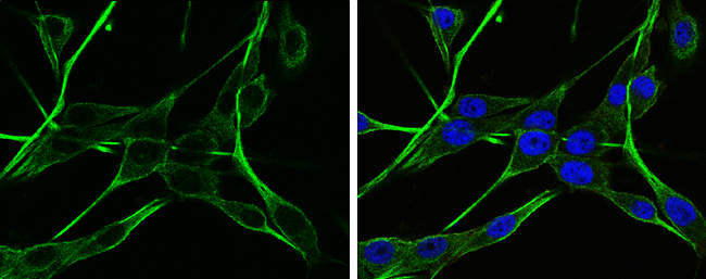
- NAGLU antibody detects NAGLU protein at lysosome by immunofluorescent analysis.Sample: HeLa cells were fixed in 4% paraformaldehyde at RT for 15 min.Green: NAGLU protein stained by NAGLU antibody (GTX105504) diluted at 1:500.Blue: Hoechst 33342 staining.Scale bar = 10 £gm.

- Submitted by
- GeneTex (provider)
- Main image

- Experimental details
- NAGLU antibody detects NAGLU protein at lysosome by immunofluorescent analysis.Sample: HeLa cells were fixed in 4% paraformaldehyde at RT for 15 min.Green: NAGLU protein stained by NAGLU antibody (GTX105504) diluted at 1:500.Blue: Hoechst 33342 staining.

- Submitted by
- GeneTex (provider)
- Main image
- Experimental details
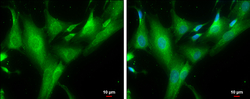
- NAGLU antibody detects NAGLU protein at cytoplasm by immunofluorescent analysis.Sample: SK-N-SH cells were fixed in ice-cold MeOH for 5 min.Green: NAGLU protein stained by NAGLU antibody (GTX105504) diluted at 1:500.Blue: Hoechst 33342 staining.Scale bar = 10 £gm.

Supportive validation
- Submitted by
- GeneTex (provider)
- Main image
- Experimental details
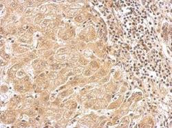
- NAGLU antibody detects NAGLU protein at cytosol on human hepatoma by immunohistochemical analysis. Sample: Paraffin-embedded hepatoma. NAGLU antibody (GTX105504) dilution: 1:500.

- Submitted by
- GeneTex (provider)
- Main image

- Experimental details
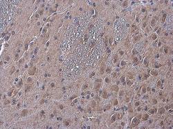
- NAGLU antibody detects NAGLU protein at cytoplasm in mouse brain by immunohistochemical analysis. Sample: Paraffin-embedded mouse brain. NAGLU antibody (GTX105504) diluted at 1:500.

- Submitted by
- GeneTex (provider)
- Main image

- Experimental details
- NAGLU antibody detects NAGLU protein at cytoplasm in rat brain by immunohistochemical analysis. Sample: Paraffin-embedded rat brain. Green: NAGLU antibody (GTX105504) diluted at 1:200. The signal was developed using goat anti-rabbit IgG antibody (Dylight488) (GTX213110-04).Blue: Nuclear staining with Hoechst 33342.

- Submitted by
- GeneTex (provider)
- Main image

- Experimental details
- NAGLU antibody detects NAGLU protein at cytoplasm in mouse pancreas by immunohistochemical analysis. Sample: Paraffin-embedded mouse pancreas. NAGLU antibody (GTX105504) diluted at 1:500.

- Submitted by
- GeneTex (provider)
- Main image

- Experimental details
- NAGLU antibody detects NAGLU protein at cytoplasm in rat pancreas by immunohistochemical analysis. Sample: Paraffin-embedded rat pancreas. NAGLU antibody (GTX105504) diluted at 1:500.

- Submitted by
- GeneTex (provider)
- Main image

- Experimental details
- NAGLU antibody detects NAGLU Protein expression by immunohistochemical analysis.Sample: Frozen-sectioned adult mouse cerebellum. Green: NAGLU stained by NAGLU antibody (GTX105504) diluted at 1:250.Red: NF-H, stained by NF-H antibody [GT114] (GTX634289) diluted at 1:500.Blue: Fluoroshield with DAPI (GTX30920).

- Submitted by
- GeneTex (provider)
- Main image

- Experimental details
- NAGLU antibody detects NAGLU protein at cytoplasm in mouse brain by immunohistochemical analysis. Sample: Paraffin-embedded mouse brain. NAGLU antibody (GTX105504) diluted at 1:500.

- Submitted by
- GeneTex (provider)
- Main image

- Experimental details
- NAGLU antibody detects NAGLU protein at cytoplasm in rat duodenum by immunohistochemical analysis. Sample: Paraffin-embedded rat duodenum. NAGLU antibody (GTX105504) diluted at 1:500.

- Submitted by
- GeneTex (provider)
- Main image
- Experimental details
- NAGLU antibody detects NAGLU protein at cytoplasm in mouse brain by immunohistochemical analysis. Sample: Paraffin-embedded mouse brain. NAGLU antibody (GTX105504) diluted at 1:250.

- Submitted by
- GeneTex (provider)
- Main image

- Experimental details
- NAGLU antibody detects NAGLU protein at cytoplasm in rat duodenum by immunohistochemical analysis. Sample: Paraffin-embedded rat duodenum. NAGLU antibody (GTX105504) diluted at 1:250.
