 Explore
 Validate
 Learn
Learn Western blot
Antibody data
- Antibody Data
- Antigen structure
- References [2]
- Comments [0]
- Validations
- Western blot [1]
- Immunohistochemistry [1]
Submit
Validation data
Reference
Comment
Report error
- Product number
- NBP1-59337 - Provider product page

- Provider
- Novus Biologicals
- Proper citation
- Novus Cat#NBP1-59337, RRID:AB_11025903
- Product name
- Rabbit Polyclonal Hepcidin Antimicrobial Peptide Antibody
- Antibody type
- Polyclonal
- Description
- Immunogen affinity purified.
- Reactivity
- Human, Mouse, Bovine
- Host
- Rabbit
- Isotype
- IgG
- Vial size
- 100 ul
- Storage
- Store at 4C short term. Aliquot and store at -20C long term. Avoid freeze-thaw cycles.
Submitted references Local synthesis of hepcidin in the anterior segment of the eye: A novel observation with physiological and pathological implications.
Evidence Suggesting a Role of Iron in a Mouse Model of Nephrogenic Systemic Fibrosis.
Ashok A, Chaudhary S, McDonald D, Kritikos A, Bhargava D, Singh N
Experimental eye research 2020 Jan;190:107890
Experimental eye research 2020 Jan;190:107890
Evidence Suggesting a Role of Iron in a Mouse Model of Nephrogenic Systemic Fibrosis.
Bose C, Megyesi JK, Shah SV, Hiatt KM, Hall KA, Karaduta O, Swaminathan S
PloS one 2015;10(8):e0136563
PloS one 2015;10(8):e0136563
No comments: Submit comment
Supportive validation
- Submitted by
- Novus Biologicals (provider)
- Main image
- Experimental details
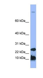
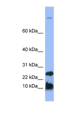
- Western Blot: Hepcidin Antimicrobial Peptide Antibody [NBP1-59337] - Human Spleen lysate, concentration 0.2-1 ug/ml.
Supportive validation
- Submitted by
- Novus Biologicals (provider)
- Main image
- Experimental details
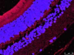
- Immunohistochemistry-Paraffin: Hepcidin Antimicrobial Peptide Antibody [NBP1-59337] - Paraffin embedded sections of posterior segment of mouse eye, red is Hepcidin and blue is Hoeschst. 4um sections were rehydrated with xylene, followed by a decreasing ethanol concentration gradient (100%, 90%, 70%) and a wash with diH2O. Heat-mediated antigen retrieval performed using EDTA buffer (10mM Trizma Base, 1mM EDTA solution, 0.05% Tween 20, pH 9.0) in an autoclave for 30min. Primary antibody against Hepcidin was diluted 1:10, in blocking solution containing 0.1% BSA, 0.05% Triton X-100, and 5% normal donkey serum in TBS. Sections were incubated for 48hrs at room temperature and then 24hrs at 4C. Tissues were washed with TBS-T (6x5min), and immunoreactivity for Hep was developed. Image from verified customer review.
