 Explore
 Validate
 Learn
Learn Western blot
Antibody data
- Antibody Data
- Antigen structure
- References [15]
- Comments [0]
- Validations
- Western blot [7]
- Immunocytochemistry [2]
- Other assay [9]
Submit
Validation data
Reference
Comment
Report error
- Product number
- MA1-16757 - Provider product page

- Provider
- Invitrogen Antibodies
- Product name
- GAPDH Monoclonal Antibody (1D4)
- Antibody type
- Monoclonal
- Antigen
- Purifed from natural sources
- Description
- This antibody is likely to react with most mammals.
- Antibody clone number
- 1D4
- Concentration
- 1 mg/mL
Submitted references Givinostat-Liposomes: Anti-Tumor Effect on 2D and 3D Glioblastoma Models and Pharmacokinetics.
An in vivo Cell-Based Delivery Platform for Zinc Finger Artificial Transcription Factors in Pre-clinical Animal Models.
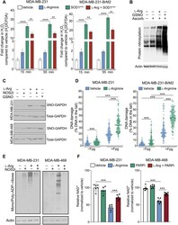
The metabolic adaptation evoked by arginine enhances the effect of radiation in brain metastases.
LncRNA HOXA11-AS aggravates the keloid formation by targeting miR-148b-3p/IGFBP5 axis.
Evaluation of β-Catenin Inhibition of Axitinib and Nitazoxanide in Human Monocyte-Derived Dendritic Cells.
Mitochondrial-Derived Vesicles Protect Cardiomyocytes Against Hypoxic Damage.
The regulation of Oct4 in human gingival fibroblasts stimulated by cyclosporine A: Preliminary observations.
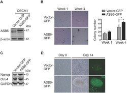
ASB6 Promotes the Stemness Properties and Sustains Metastatic Potential of Oral Squamous Cell Carcinoma Cells by Attenuating ER Stress.
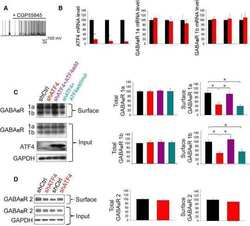
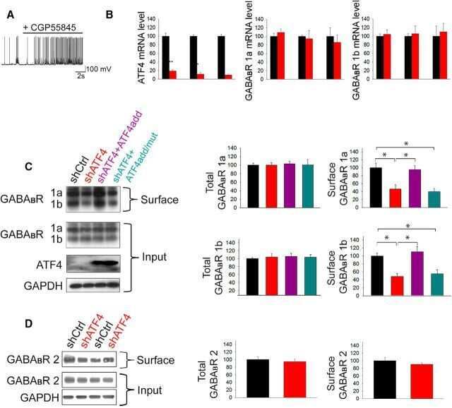
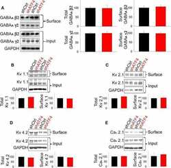
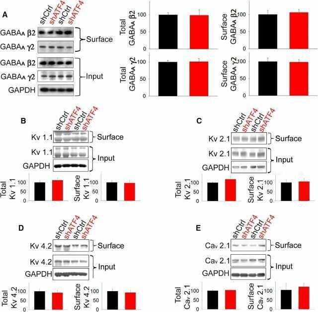
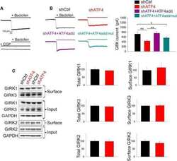
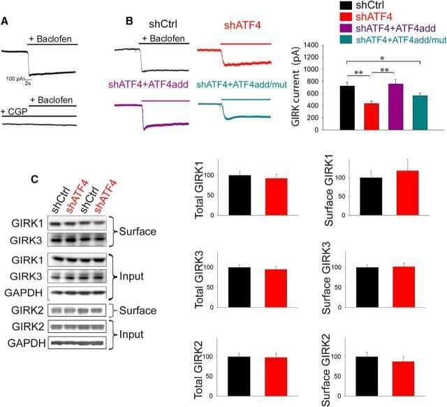
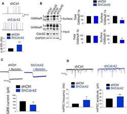
Activating Transcription Factor 4 (ATF4) Regulates Neuronal Activity by Controlling GABA(B)R Trafficking.
CREB overexpression in dorsal CA1 ameliorates long-term memory deficits in aged rats.
Emodin suppresses silica-induced lung fibrosis by promoting Sirt1 signaling via direct contact.
Epigenetic dysregulation of KCa 3.1 channels induces poor prognosis in lung cancer.
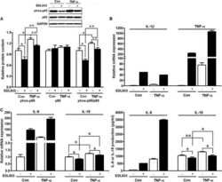
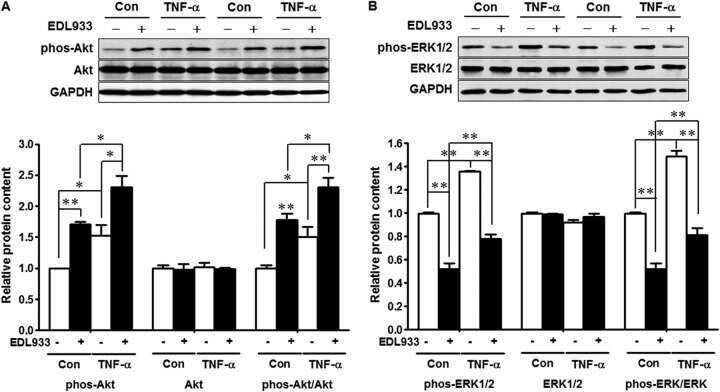
Host inflammatory response inhibits Escherichia coli O157:H7 adhesion to gut epithelium through augmentation of mucin expression.
Disease sequence from mutant rhodopsin allele to rod and cone photoreceptor degeneration in man.
Antigen-antibody interaction. Synthetic peptides define linear antigenic determinants recognized by monoclonal antibodies directed to the cytoplasmic carboxyl terminus of rhodopsin.
Taiarol L, Bigogno C, Sesana S, Kravicz M, Viale F, Pozzi E, Monza L, Carozzi VA, Meregalli C, Valtorta S, Moresco RM, Koch M, Barbugian F, Russo L, Dondio G, Steinkühler C, Re F
Cancers 2022 Jun 16;14(12)
Cancers 2022 Jun 16;14(12)
An in vivo Cell-Based Delivery Platform for Zinc Finger Artificial Transcription Factors in Pre-clinical Animal Models.
Deng P, Halmai JANM, Beitnere U, Cameron D, Martinez ML, Lee CC, Waldo JJ, Thongphanh K, Adhikari A, Copping N, Petkova SP, Lee RD, Lock S, Palomares M, O'Geen H, Carter J, Gonzalez CE, Buchanan FKB, Anderson JD, Fierro FA, Nolta JA, Tarantal AF, Silverman JL, Segal DJ, Fink KD
Frontiers in molecular neuroscience 2021;14:789913
Frontiers in molecular neuroscience 2021;14:789913
The metabolic adaptation evoked by arginine enhances the effect of radiation in brain metastases.
Marullo R, Castro M, Yomtoubian S, Calvo-Vidal MN, Revuelta MV, Krumsiek J, Cho A, Morgado PC, Yang S, Medina V, Roth BM, Bonomi M, Keshari KR, Mittal V, Navigante A, Cerchietti L
Science advances 2021 Nov 5;7(45):eabg1964
Science advances 2021 Nov 5;7(45):eabg1964
LncRNA HOXA11-AS aggravates the keloid formation by targeting miR-148b-3p/IGFBP5 axis.
Wang J, Shen J
Biochemical and biophysical research communications 2021 Dec 3;581:60-67
Biochemical and biophysical research communications 2021 Dec 3;581:60-67
Evaluation of β-Catenin Inhibition of Axitinib and Nitazoxanide in Human Monocyte-Derived Dendritic Cells.
Azeem W, Bakke RM, Gabriel B, Appel S, Øyan AM, Kalland KH
Biomedicines 2021 Aug 3;9(8)
Biomedicines 2021 Aug 3;9(8)
Mitochondrial-Derived Vesicles Protect Cardiomyocytes Against Hypoxic Damage.
Li B, Zhao H, Wu Y, Zhu Y, Zhang J, Yang G, Yan Q, Li J, Li T, Liu L
Frontiers in cell and developmental biology 2020;8:214
Frontiers in cell and developmental biology 2020;8:214
The regulation of Oct4 in human gingival fibroblasts stimulated by cyclosporine A: Preliminary observations.
Yu CC, Liu CM, Lin TC, Su NY, Yang LC, Chang YC
Journal of dental sciences 2020 Jun;15(2):176-180
Journal of dental sciences 2020 Jun;15(2):176-180
ASB6 Promotes the Stemness Properties and Sustains Metastatic Potential of Oral Squamous Cell Carcinoma Cells by Attenuating ER Stress.
Hung KF, Liao PC, Chen CK, Chiu YT, Cheng DH, Kawasumi M, Kao SY, Lo JF
International journal of biological sciences 2019;15(5):1080-1090
International journal of biological sciences 2019;15(5):1080-1090
Activating Transcription Factor 4 (ATF4) Regulates Neuronal Activity by Controlling GABA(B)R Trafficking.
Corona C, Pasini S, Liu J, Amar F, Greene LA, Shelanski ML
The Journal of neuroscience : the official journal of the Society for Neuroscience 2018 Jul 4;38(27):6102-6113
The Journal of neuroscience : the official journal of the Society for Neuroscience 2018 Jul 4;38(27):6102-6113
CREB overexpression in dorsal CA1 ameliorates long-term memory deficits in aged rats.
Yu XW, Curlik DM, Oh MM, Yin JC, Disterhoft JF
eLife 2017 Jan 4;6
eLife 2017 Jan 4;6
Emodin suppresses silica-induced lung fibrosis by promoting Sirt1 signaling via direct contact.
Yang T, Wang J, Pang Y, Dang X, Ren H, Liu Y, Chen M, Shang D
Molecular medicine reports 2016 Nov;14(5):4643-4649
Molecular medicine reports 2016 Nov;14(5):4643-4649
Epigenetic dysregulation of KCa 3.1 channels induces poor prognosis in lung cancer.
Bulk E, Ay AS, Hammadi M, Ouadid-Ahidouch H, Schelhaas S, Hascher A, Rohde C, Thoennissen NH, Wiewrodt R, Schmidt E, Marra A, Hillejan L, Jacobs AH, Klein HU, Dugas M, Berdel WE, Müller-Tidow C, Schwab A
International journal of cancer 2015 Sep 15;137(6):1306-17
International journal of cancer 2015 Sep 15;137(6):1306-17
Host inflammatory response inhibits Escherichia coli O157:H7 adhesion to gut epithelium through augmentation of mucin expression.
Xue Y, Zhang H, Wang H, Hu J, Du M, Zhu MJ
Infection and immunity 2014 May;82(5):1921-30
Infection and immunity 2014 May;82(5):1921-30
Disease sequence from mutant rhodopsin allele to rod and cone photoreceptor degeneration in man.
Cideciyan AV, Hood DC, Huang Y, Banin E, Li ZY, Stone EM, Milam AH, Jacobson SG
Proceedings of the National Academy of Sciences of the United States of America 1998 Jun 9;95(12):7103-8
Proceedings of the National Academy of Sciences of the United States of America 1998 Jun 9;95(12):7103-8
Antigen-antibody interaction. Synthetic peptides define linear antigenic determinants recognized by monoclonal antibodies directed to the cytoplasmic carboxyl terminus of rhodopsin.
Hodges RS, Heaton RJ, Parker JM, Molday L, Molday RS
The Journal of biological chemistry 1988 Aug 25;263(24):11768-75
The Journal of biological chemistry 1988 Aug 25;263(24):11768-75
No comments: Submit comment
Supportive validation
- Submitted by
- Invitrogen Antibodies (provider)
- Main image

- Experimental details
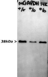
- Sciatic nerves of mouse wild type (+/+), heterozygous (+/o) and homozygous (o/o) for knock out of peripheral myelin protein 21 (pmp21) were homogenized in SDS-PAGE sample buffer and run out for western blots. Blots were probed with MA1-16757 mouse monoclonal antibody to glyceraldehyde 3-phosphate dehydrogenase (GAPDH). Antibody was used at dilution of 1:1,000. Signal was revealed in a few seconds with chemiluminescence, indicating that lower antibody concentrations would also have worked well. Note the sharp clear band at 38kDa, the expected molecular weight for GAPDH.
- Submitted by
- Invitrogen Antibodies (provider)
- Main image
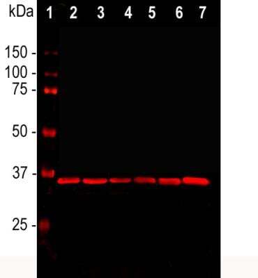
- Experimental details
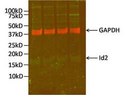
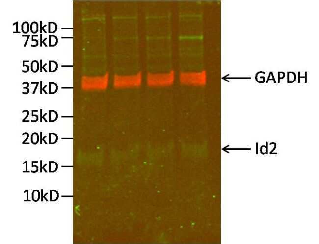
- Western blot analysis of GAPDH was performed by loading 10 µg of Id2-transfected SK-N-BE human neuroblastoma cells (in replicate) per well onto an SDS-PAGE gel. Proteins were transferred to a PVDF membrane and blocked for 1 hour. The membrane was probed with a GAPDH monoclonal antibody (Product # MA5-16757) at a dilution of 1:2000 overnight at 4ºC, washed in 0.1% PBS-Tween, and probed with an infrared dye-conjugated anti-mouse IgG secondary antibody (shown in red) at a dilution of 1:20,000 for 1 hour. The blot was also probed with an Id2 monoclonal antibody (Product # MA5-14777) at a dilution of 1:500 followed by an infrared Dye-conjugated anti-rabbit IgG secondary antibody (shown in green) at a dilution of 1:15,000. Detection was performed using a fluorescent scanner. Data courtesy of the Innovators Program.
- Submitted by
- Invitrogen Antibodies (provider)
- Main image
- Experimental details
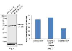
- Knockdown of GAPDH was achieved by transfecting HeLa cells with GAPDH specific validated siRNAs (Silencer® select Product # s13960 ). Western blot analysis (Fig. a) was performed using whole cell extracts from the GAPDH knockdown cells (lane 3), non-specific scrambled siRNA transfected cells (lane 2) and untransfected cells (lane 1). The blots were probed with GAPDH Monoclonal Antibody (Product # MA1-16757, 1:2000 dilution) and Goat anti-Mouse IgG (H+L) Superclonal™ Secondary Antibody, HRP conjugate (Product # A28177, 0.25 µg/mL, 1:4000 dilution). Densitometric analysis of this western blot is shown in histogram (Fig. b). Decrease in signal upon siRNA mediated knock down confirms that antibody is specific to GAPDH.

- Submitted by
- Invitrogen Antibodies (provider)
- Main image

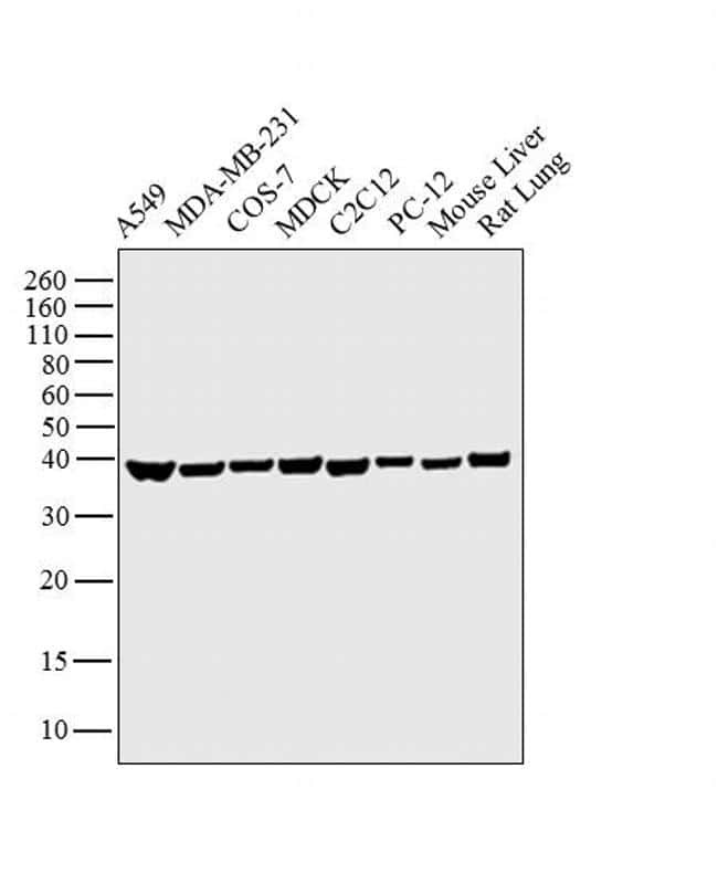
- Experimental details
- Western blot analysis was performed on whole cell extracts (30 µg lysate) of A549 (Lane 1), MDA-MB-231 (Lane 2), COS-7 (Lane 3), MDCK (Lane 4), C2C12 (Lane 5), PC-12 (Lane 6) and tissue extracts (30 µg lysate) of Mouse Liver (Lane 7) and Rat Lung (Lane 8 ). The blot was probed with Anti-GAPDH Monoclonal Antibody (Product # MA1-16757, 1:1000 dilution) and detected by chemiluminescence using Goat anti-Mouse IgG (H+L) Superclonal™ Secondary Antibody, HRP conjugate (Product # A28177, 0.25 µg/mL, 1:4000 dilution). A 37 kDa band corresponding to GAPDH was observed across the cell lines and tissue extracts tested.
- Submitted by
- Invitrogen Antibodies (provider)
- Main image
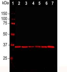
- Experimental details
- Western blot analysis of GAPDH in cell line lysates. Samples were incubated in GAPDH monoclonal antibody (Product # MA1-16757 using a dilution of 1:2,000. [1] protein standard, [2] HEK293, [3] HeLa, [4] SH-SY5Y, [5] COS1, [6] NIH-3T3, and [7] C6 cells. The GAPDH antibody reveals a single band at ~37 kDa in all cell lines. GAPDH is a house keeping protein, the level of which is relatively unaffected by most experimental manipulations, and, as a result, this antibody has been widely used as a ting loading control.
- Submitted by
- Invitrogen Antibodies (provider)
- Main image
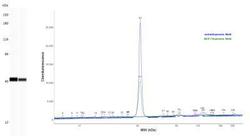
- Experimental details
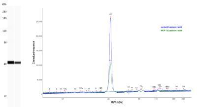
- Western blot analysis of GAPDH in 0.05 mg/mL Jurkat and MCF-7 cell lysates. Samples were incubated in GAPDH monoclonal antibody (Product # MA1-16757) using a dilution of 1:25. Electropherogram image of corresponding Simple Western lanes view at WES molecular weight of 40 kDa.
- Submitted by
- Invitrogen Antibodies (provider)
- Main image

- Experimental details
- Western blot analysis was performed on whole cell extracts (30 µg lysate) of A549 (Lane 1), MDA-MB-231 (Lane 2), COS-7 (Lane 3), MDCK (Lane 4), C2C12 (Lane 5), PC-12 (Lane 6) and tissue extracts (30 µg lysate) of Mouse Liver (Lane 7) and Rat Lung (Lane 8 ). The blot was probed with Anti-GAPDH Monoclonal Antibody (Product # MA1-16757, 1:1000 dilution) and detected by chemiluminescence using Goat anti-Mouse IgG (H+L) Superclonal™ Secondary Antibody, HRP conjugate (Product # A28177, 0.25 µg/mL, 1:4000 dilution). A 37 kDa band corresponding to GAPDH was observed across the cell lines and tissue extracts tested.

Supportive validation
- Submitted by
- Invitrogen Antibodies (provider)
- Main image
- Experimental details
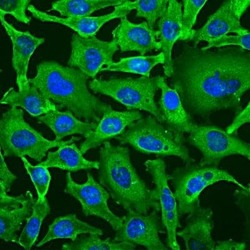
- Immunocytochemistry analysis of GAPDH in HeLa cells. Samples were incubated in GAPDH monoclonal antibody (Product # MA1-16757) using a dilution of 1:100. Antibody (Green). DAPI staining of nuclear DNA (Blue). The GAPDHantibody produces strong cytoplasmic staining of healthy cells.

- Submitted by
- Invitrogen Antibodies (provider)
- Main image

- Experimental details
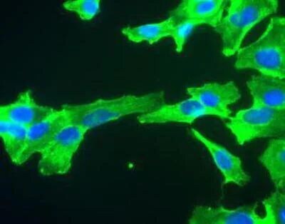
- Immunocytochemistry analysis of GAPDH in SH-SY5Y cells. Samples were incubated in GAPDH monoclonal antibody (Product # MA1-16757). GAPDH antibody (green). Nuclear DNA is stained with Hoechst dye (blue).
Supportive validation
- Submitted by
- Invitrogen Antibodies (provider)
- Main image
- Experimental details
- NULL

- Submitted by
- Invitrogen Antibodies (provider)
- Main image
- Experimental details
- NULL

- Submitted by
- Invitrogen Antibodies (provider)
- Main image

- Experimental details
- NULL
- Submitted by
- Invitrogen Antibodies (provider)
- Main image
- Experimental details
- NULL
- Submitted by
- Invitrogen Antibodies (provider)
- Main image
- Experimental details
- NULL
- Submitted by
- Invitrogen Antibodies (provider)
- Main image
- Experimental details
- NULL
- Submitted by
- Invitrogen Antibodies (provider)
- Main image
- Experimental details
- NULL

- Submitted by
- Invitrogen Antibodies (provider)
- Main image
- Experimental details
- Fig. 5. l -Arginine generates peroxynitrate, GAPDH nitrosylation, and protein ADP-ribosylation. ( A ) Peroxynitrate levels in MDA-MB-231 and MDA-MB-231-BrM2 cells treated with vehicle, l -arginine, the SOD mimetic tempol (SOD1 mim ), or the combination of l -arginine and SOD1 mim for 15 and 55 min. ( B ) Total protein nitrosylation in MDA-MB-231 cells exposed to l -arginine for 60 min. GSNO was used as a positive control. ( C ) SNO-GAPDH (and total GAPDH) in MDA-MB-231 and MDA-MB-468 cells exposed for 60 min to l -arginine alone or in combination with the NOS2 inhibitor 1400W. GSNO was used as a positive control. ( D ) DNA damage levels assessed by comet assay in MDA-MB-231 and MDA-MB-231-BrM2 cells exposed to vehicle or l -arginine for 60 min with and without Fpg. ( E ) Total mono- and poly-ADP-ribosylated proteins in MDA-MB-231 and MDA-MB-468 cells exposed to l -arginine alone or in combination with the NOS2 inhibitor 1400W. ( F ) NAD + levels in MDA-MB-231 and MDA-MB-468 cells exposed to l -arginine alone or in combination with the PARP inhibitor olaparib. ** P < 0.001 and *** P < 0.0001.

- Submitted by
- Invitrogen Antibodies (provider)
- Main image
- Experimental details
- Figure 1 The effect of ASB6 overexpression on soft agar colony formation, stemness genes expression, and tumor sphere formation of OSCC cells. The OECM1 cells with stable overexpression of green fluorescent protein (vector GFP) or GFP-tagged ASB6 (ASB6-GFP) were validated by western blot for ASB6 (with as beta-actin as the loading control) (A) , and were subjected to anchorage-independent growth analysis by the soft agar assay (B) , western blots analysis for Nanog and Oct-4 (with as the GAPDH as loading control) (C) , and tumor sphere formation analysis (D) .* P < 0.05.
