Explore
 Validate
 Learn
Learn Western blot
Antibody data
- Antibody Data
- Antigen structure
- References [0]
- Comments [0]
- Validations
- Western blot [2]
- Immunocytochemistry [2]
- Immunohistochemistry [2]
Submit
Validation data
Reference
Comment
Report error
- Product number
- PA5-30485 - Provider product page

- Provider
- Invitrogen Antibodies
- Product name
- EDC4 Polyclonal Antibody
- Antibody type
- Polyclonal
- Antigen
- Recombinant protein fragment
- Description
- Recommended positive controls: NT2D1, IMR32, U87-MG, MCF-7, mouse brain.
- Concentration
- 1 mg/mL
No comments: Submit comment
Supportive validation
- Submitted by
- Invitrogen Antibodies (provider)
- Main image

- Experimental details
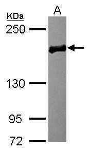
- Western Blot using EDC4 Polyclonal Antibody (Product # PA5-30485). Sample (30 µg of whole cell lysate). Lane A: MCF-7. 5% SDS PAGE. EDC4 Polyclonal Antibody (Product # PA5-30485) diluted at 1:3,000.
- Submitted by
- Invitrogen Antibodies (provider)
- Main image

- Experimental details
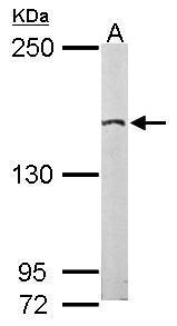
- Western Blot using EDC4 Polyclonal Antibody (Product # PA5-30485). Sample (50 µg of whole cell lysate). Lane A: mouse brain . 5% SDS PAGE. EDC4 Polyclonal Antibody (Product # PA5-30485) diluted at 1:2,000.
Supportive validation
- Submitted by
- Invitrogen Antibodies (provider)
- Main image
- Experimental details
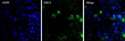
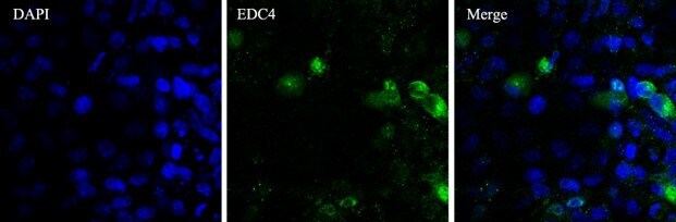
- Immunofluorescent analysis of EDC4 (green) in Hela cells. The cells were fixed with 4% paraformaldehyde for 15 minutes, permeabilized with 0.1% Triton X-100 in PBS for 15 minutes, and blocked with 2% BSA in PBS for 60 minutes at room temperature. Cells were stained with a EDC4 polyclonal antibody (Product # PA5-30485) at a dilution of 2 µg/mL in staining buffer for 3 hour at room temperature, and then incubated with a Goat anti-rabbit IgG Secondary Antibody, Alexa Fluor 488 conjugate (Product # A32731) at a dilution of 1:1000 for 1 hour at room temperature (green). Nuclei (blue) were counterstained with DAPI dye. Images were taken at 100X magnification. Data courtesy of Thermo Scientific KOL Program.
- Submitted by
- Invitrogen Antibodies (provider)
- Main image
- Experimental details
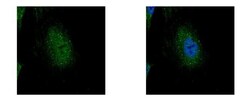
- EDC4 Polyclonal Antibody detects EDC4 protein at P-body by confocal immunofluorescent analysis. Sample: HeLa cells were fixed in 4% paraformaldehyde at RT for 15 min. Green: EDC4 protein stained by EDC4 Polyclonal Antibody (Product # PA5-30485) diluted at 1:500. Blue: Hoechst 33343 staining. [Images captured by Olympus FV10i Confocal Laser Scanning Microscope].

Supportive validation
- Submitted by
- Invitrogen Antibodies (provider)
- Main image
- Experimental details
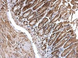
- EDC4 Polyclonal Antibody detects EDC4 protein at cytosol and nucleus on mouse duodenum by immunohistochemical analysis. Sample: Paraffin-embedded mouse duodenum. EDC4 Polyclonal Antibody (Product # PA5-30485) dilution: 1:500. Antigen Retrieval: EDTA based buffer, pH 8.0, 15 min.

- Submitted by
- Invitrogen Antibodies (provider)
- Main image
- Experimental details
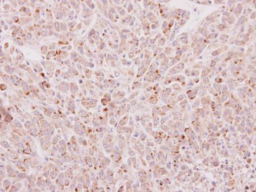
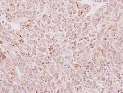
- Immunohistochemical analysis of paraffin-embedded U87 xenograft, using EDC4 (Product # PA5-30485) antibody at 1:100 dilution. Antigen Retrieval: EDTA based buffer, pH 8.0, 15 min.