 Explore
 Validate
 Learn
LearnPA5-23043
antibody from Invitrogen Antibodies
Targeting: CCR2
CC-CKR-2, CD192, CKR2, CMKBR2, FLJ78302, MCP-1-R
 Immunocytochemistry
 Immunohistochemistry
Antibody data
- Antibody Data
- Antigen structure
- References [2]
- Comments [0]
- Validations
- Immunocytochemistry [2]
- Flow cytometry [3]
- Other assay [3]
Submit
Validation data
Reference
Comment
Report error
- Product number
- PA5-23043 - Provider product page

- Provider
- Invitrogen Antibodies
- Product name
- CCR2 Polyclonal Antibody
- Antibody type
- Polyclonal
- Antigen
- Synthetic peptide
- Reactivity
- Human, Mouse
- Host
- Rabbit
- Isotype
- IgG
- Vial size
- 100 µL
- Concentration
- 1 mg/mL
- Storage
- -20° C, Avoid Freeze/Thaw Cycles
Submitted references TCF4 enhances hepatic metastasis of colorectal cancer by regulating tumor-associated macrophage via CCL2/CCR2 signaling.
Effect of CCR2 inhibitor-loaded lipid micelles on inflammatory cell migration and cardiac function after myocardial infarction.
Tu W, Gong J, Zhou Z, Tian D, Wang Z
Cell death & disease 2021 Sep 27;12(10):882
Cell death & disease 2021 Sep 27;12(10):882
Effect of CCR2 inhibitor-loaded lipid micelles on inflammatory cell migration and cardiac function after myocardial infarction.
Wang J, Seo MJ, Deci MB, Weil BR, Canty JM, Nguyen J
International journal of nanomedicine 2018;13:6441-6451
International journal of nanomedicine 2018;13:6441-6451
No comments: Submit comment
Supportive validation
- Submitted by
- Invitrogen Antibodies (provider)
- Main image
- Experimental details
- Immunofluorescent analysis of CCR2 using a polyclonal antibody (Product # PA5-23043).

- Submitted by
- Invitrogen Antibodies (provider)
- Main image

- Experimental details
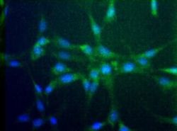
- Immunocytochemistry analysis of CCR2 in NIH/3T3 cells. Samples were incubated in CCR2 polyclonal antibody (Product # PA5-23043).

Supportive validation
- Submitted by
- Invitrogen Antibodies (provider)
- Main image

- Experimental details
- Flow cytometry analysis of CCR2 using a polyclonal antibody (Product # PA5-23043).

- Submitted by
- Invitrogen Antibodies (provider)
- Main image

- Experimental details
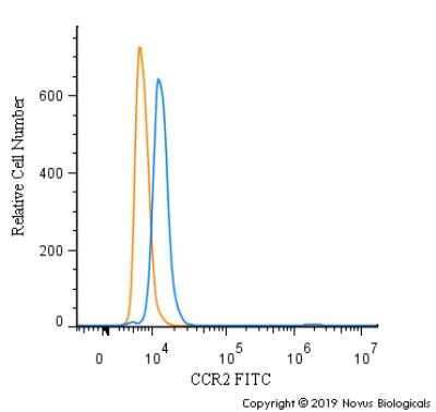
- Flow cytometry of CCR2 in THP-1 cells. Samples were incubated in CCR2 polyclonal antibody (Product # PA5-23043) using a dilution of 2.5 µg/mL for 30 minutes at room temperature. Antibody (blue) and a matched isotype control (orange). Cells were fixed with 4% PFA and then permeabilized with 0.1% saponin. Both antibodies were conjugated to DyLight 488.

- Submitted by
- Invitrogen Antibodies (provider)
- Main image

- Experimental details
- Flow cytometry of CCR2 in THP-1 cells. Samples were incubated in CCR2 polyclonal antibody (Product # PA5-23043) using a dilution of 10 µg/mL for 30 minutes at room temperature. Antibody (blue) and a matched isotype control (orange). Cells were fixed with 4% PFA and then permeabilized with 0.1% saponin. Both antibodies were conjugated to FITC.
Supportive validation
- Submitted by
- Invitrogen Antibodies (provider)
- Main image

- Experimental details
- Fig. 5 TCF4 enhances TAM recruitment and M2 polarization in the tumor through regulating the CCL2-CCR2 axis. A A heatmap of differential expression genes between MC38TCF4-sh cells compared with MC38Control cells using a chemokine PCR array. n = 4, per group. B CCL2 protein concentrations in the culture medium of MC38Control and MC38TCF4-sh cells were evaluated by ELISA. n = 4, per group. C CCL2, CCR2 and CCR4 mRNA expression in metastatic hepatic tumors of mice model received MC38Control and MC38TCF4-sh cells spleen injection. n = 8 per group. D Representative images of Immunofluorescence staining for F4/80(red), CCR2 (green) and DAPI (blue) in mouse hepatic metastases. Scale bar, 50 mum. E Quantification of F4/80 and CCR2 double positive cells per mm 2 tumor area. n = 8 per group; five fields were counted per sample. F CCL2 Immunohistochemistry staining and quantification in the human primary colorectal cancer and hepatic metastatses. Scale bar, 100 mum. Primary cancer, n = 21; hepatic metastases, n = 21; five fields were counted per sample. G Q-PCR results of CCL2, CCR2, and CCR4 mRNA expression in human primary colorectal cancer and hepatic metastatses. H Representative images of Immunofluorescence staining for CD68(red), CCR2(green) and DAPI(blue) in human primary colorectal cancer and hepatic metastatses. Scale bar, 100 mum. I Quantification of CD68 + CCR2 + cells per mm2 tumor area. Primary cancer, n = 21; hepatic metastases, n = 21; five fields were counted per sample.

- Submitted by
- Invitrogen Antibodies (provider)
- Main image

- Experimental details
- Fig. 7 Silencing of CCL2 in MC38 cells inhibits hepatic tumor growth through regulation of TAM recruitment and polarization in mouse model. A The schematic protocol of the mouse model received MC38 spleen injection. B Representative images and tumor nodule numbers of mouse liver with colorectal cancer metastases. MC38Control group, n = 8; MC38CCL2-sh group n = 7; Scale bar, 10 mm. C The liver weight of the mice was evaluated. D F4/80 Immunohistochemistry staining and quantification in the metastatic tumor region. MC38Control group, n = 8; MC38CCL2-sh group n = 7; Scale bar, 100 mum; five fields were counted per sample. E F4/80 mRNA expression in the tumor was analyzed by Q-PCR. F Q-PCR analysis of CCL2, CCR2, and CCR4 mRNA expression in the tumors. G Representative images of Immunofluorescence staining for F4/80(red), CCR2(green) and DAPI(blue) in mouse hepatic metastases. Scale bar, 50 mum. H Quantification of F4/80 and CCR2 double positive cells per mm2 tumor area. MC38Control group, n = 8; MC38CCL2-sh group n = 7; five fields were counted per sample. I mRNA expression of M1 macrophage markers in tumors. J M2 macrophage markers CD206, VEGF, CD163, IL-4, IL-13 mRNA expression in tumors. Data are means +- SEM. * P < 0.05, ** P < 0.01, *** P < 0.005, **** P < 0.001.

- Submitted by
- Invitrogen Antibodies (provider)
- Main image

- Experimental details
- Figure S2 Colocalization studies of DiD-labeled ( A ) CCR2-targeting micelles and ( B ) non-targeted micelles with CCR2-labeled inflammatory cells in the infarcted myocardium. The colocalization of CCR2-positive cells and DiD-labeled micelles is shown in white. ( C ) The PCC was used to quantify the degree of colocalization between CCR2-positive inflammatory cells and the DiD-labeled CCR2-targeting and non-targeted micelles. Notes: A PCC of 1 indicates perfect correlation, a PCC of 0 means no correlation, and a PCC of -1 means perfect inverse correlation. The CCR2-targeting micelles showed a significantly greater colocalization with CCR2-positive cells than non-targeted micelles ( P =0.0004). Three days after inducing myocardial infarction, mice were treated with DiD-labeled CCR2-targeting (n=3) and non-targeted micelles (n=3). 6 hours post administration, the hearts were removed and embedded in Tissue-Tek O.C.T. Frozen heart sections with 10 mum thickness were fixed and permeabilized with ice-cold acetone, and blocked with 5% bovine serum albumin in TBST. The sections were stained with an anti-mouse CCR2 antibody (Thermo Fisher Scientific, PA5-23043) at a 1:50 dilution for 1 hour at room temperature, followed by 45 minutes incubation with a secondary antibody labeled with Alexa Fluor 568 at 1:200 dilution. The nuclei were stained with DAPI. The analysis was performed in ImageJ using the Coloc 2 function. On average, three cryosections per heart were analyzed. For each c
