Explore
 Validate
 Learn
Learn Western blot
Antibody data
- Antibody Data
- Antigen structure
- References [0]
- Comments [0]
- Validations
- Western blot [2]
- Immunocytochemistry [1]
- Immunohistochemistry [5]
Submit
Validation data
Reference
Comment
Report error
- Product number
- HPA056273 - Provider product page

- Provider
- Atlas Antibodies
- Proper citation
- Atlas Antibodies Cat#HPA056273, RRID:AB_2683084
- Product name
- Anti-ARFGAP1
- Antibody type
- Polyclonal
- Reactivity
- Human
- Host
- Rabbit
- Conjugate
- Unconjugated
- Antigen sequence
TLPSMVHRVSGQPQSVTASSDKAFEDWLNDDLGSY
QGAQGNRYVGFGNTPPPQKKEDDFLNNAMSSLYSG
WSSFTTGASRFASAAKEGATKFGSQASQKASEL- Isotype
- IgG
- Vial size
- 100 µl
- Storage
- Store at +4°C for short term storage. Long time storage is recommended at -20°C.
No comments: Submit comment
Supportive validation
Supportive validation
- Submitted by
- Atlas Antibodies (provider)
- Enhanced method
- Independent antibody validation
- Main image

- Experimental details
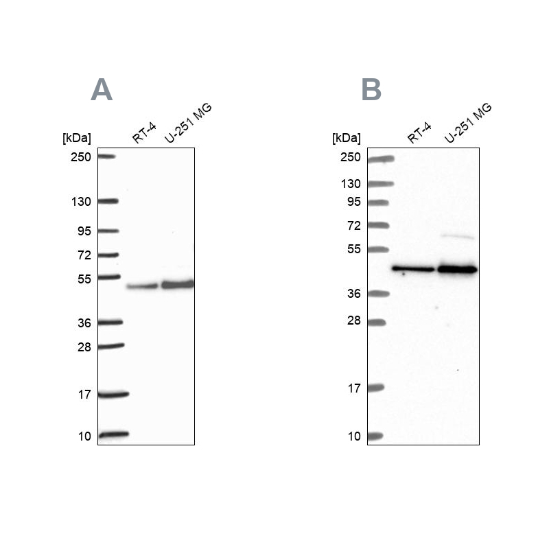
- Western blot analysis using Anti-ARFGAP1 antibody HPA056273 (A) shows similar pattern to independent antibody HPA051019 (B).
Supportive validation
- Submitted by
- Atlas Antibodies (provider)
- Main image
- Experimental details
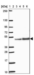
- Lane 1: Marker [kDa] 250, 130, 95, 72, 55, 36, 28, 17, 10Lane 2: Human cell line RT-4Lane 3: Human cell line U-251 MGLane 4: Human plasmaLane 5: Human Liver tissueLane 6: Human Tonsil tissue

Supportive validation
- Submitted by
- Atlas Antibodies (provider)
- Main image
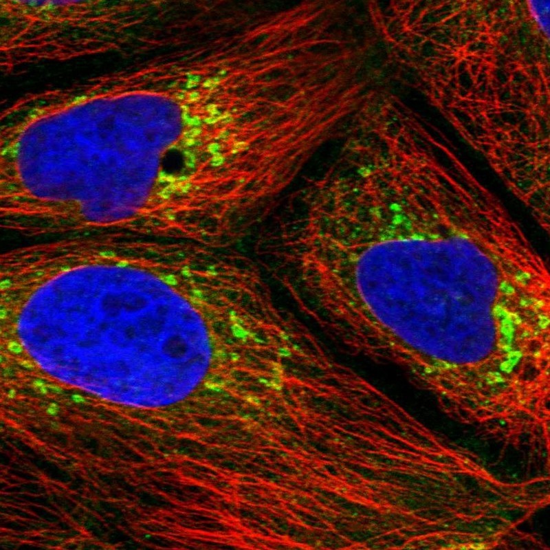
- Experimental details
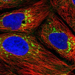
- Immunofluorescent staining of human cell line CACO-2 shows localization to cytosol, the Golgi apparatus & vesicles.
- Sample type
- HUMAN
Supportive validation
- Submitted by
- Atlas Antibodies (provider)
- Main image
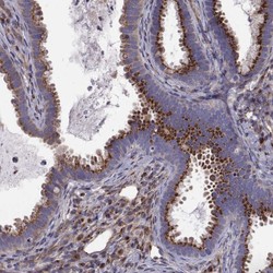
- Experimental details
- Immunohistochemical staining of human cervix, uterine shows moderate cytoplasmic positivity in glandular cells.
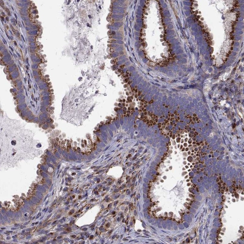
- Submitted by
- Atlas Antibodies (provider)
- Main image
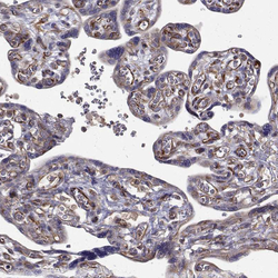
- Experimental details
- Immunohistochemical staining of human placenta shows strong cytoplasmic positivity in trophoblastic cells.
- Sample type
- HUMAN

- Submitted by
- Atlas Antibodies (provider)
- Main image
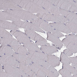
- Experimental details
- Immunohistochemical staining of human skeletal muscle shows no positivity in myocytes as expected.
- Sample type
- HUMAN

- Submitted by
- Atlas Antibodies (provider)
- Main image
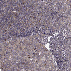
- Experimental details
- Immunohistochemical staining of human tonsil shows moderate cytoplasmic positivity in germinal center cells.
- Sample type
- HUMAN

- Submitted by
- Atlas Antibodies (provider)
- Main image

- Experimental details
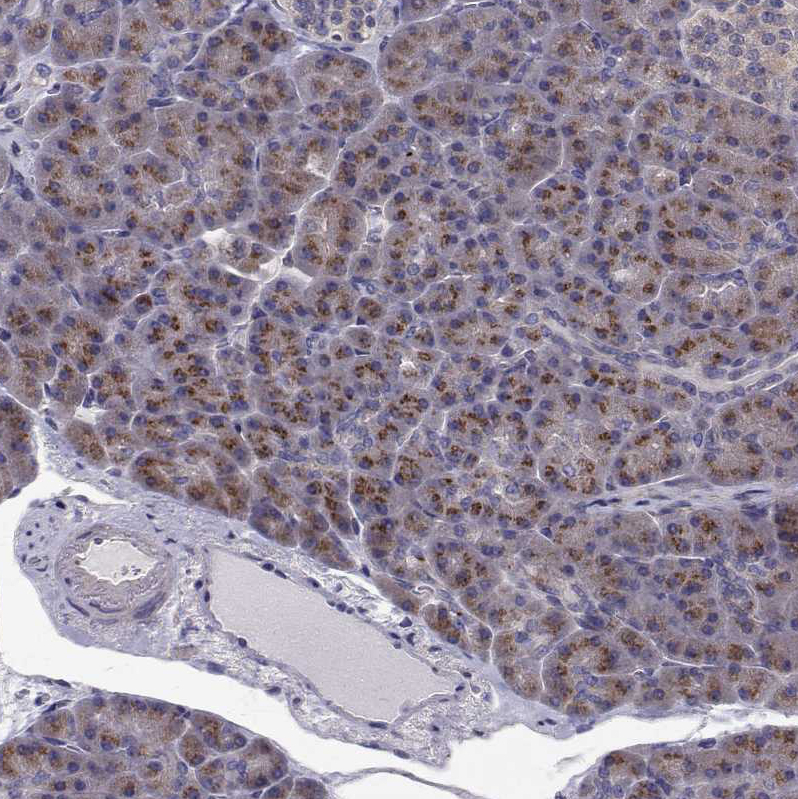
- Immunohistochemical staining of human pancreas shows strong cytoplasm granular positivity in exocrine glandular cells.
- Sample type
- HUMAN