 Explore
 Validate
 Learn
Learn Immunocytochemistry
 Immunohistochemistry
Antibody data
- Antibody Data
- Antigen structure
- References [0]
- Comments [0]
- Validations
- Immunohistochemistry [2]
- Flow cytometry [1]
Submit
Validation data
Reference
Comment
Report error
- Product number
- NB100-64347 - Provider product page

- Provider
- Novus Biologicals
- Proper citation
- Novus Cat#NB100-64347, RRID:AB_962641
- Product name
- Mouse Monoclonal CD79A Antibody
- Antibody type
- Monoclonal
- Description
- Protein G purified. NB100-64347 recognizes an epitope within the cytoplasmic domain of CD79a. CD79a, also known as mb-1, is a 45kD protein that is expressed by B lymphocytes during differentiation from early pre-B cell stage through to plasma cells. The CD79a molecule associates with CD79b (B29) to form a heterodimer that is non-covalently linked to surface immunoglobulin, forming the B-cell receptor (BCR) complex. The CD79a/CD79b heterodimers are also necessary for intracellular signalling following antigen-binding to surface immunoglobulin. Clone HM57 has been reported to work in western blotting applications.
- Reactivity
- Human, Mouse, Rat, Bovine, Canine, Chicken/Avian, Goat, Guinea Pig, Porcine, Rabbit, Simian
- Host
- Mouse
- Isotype
- IgG
- Vial size
- 0.1 mg
- Concentration
- 1.0 mg/ml
- Storage
- Store at 4C short term. Aliquot and store at -20C long term. Avoid freeze-thaw cycles.
No comments: Submit comment
Supportive validation
- Submitted by
- Novus Biologicals (provider)
- Main image
- Experimental details
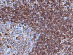
- Immunohistochemistry-Paraffin: CD79A Antibody (HM57) [NB100-64347] - Embedded human tonsil.

- Submitted by
- Novus Biologicals (provider)
- Main image
- Experimental details
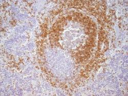
- Immunohistochemistry: CD79A Antibody (HM57) [NB100-64347] - Paraffin-embedded alcohol fixed Rat Spleen tissue (20x). Antigen retrieval pH6. Dilution: 1:500 and ON incubation at 4C. This image was submitted via customer revies.

Supportive validation
- Submitted by
- Novus Biologicals (provider)
- Main image

- Experimental details
- Flow Cytometry: CD79A Antibody (HM57) [NB100-64347] - Using the Allophycocyanin direct conjugate An intracellular stain was performed on Daudi cells with CD79A (HM 57) antibody NB100-64347APC (blue) and a matched isotype control NBP1-97005APC (orange). Cells were fixed with 4% PFA and then permeablized with 0.1% saponin. Cells were incubated in an antibody dilution of 0.5 ug/mL for 30 minutes at room temperature. Both antibodies were conjugated to Allophycocyanin.
