 Explore
 Validate
 Learn
Learn Western blot
Antibody data
- Antibody Data
- Antigen structure
- References [2]
- Comments [0]
- Validations
- Western blot [1]
- Immunohistochemistry [2]
- Other assay [1]
Submit
Validation data
Reference
Comment
Report error
- Product number
- MA1-517 - Provider product page

- Provider
- Invitrogen Antibodies
- Product name
- Mist1 Monoclonal Antibody (6E8/A12/C11P1)
- Antibody type
- Monoclonal
- Antigen
- Other
- Description
- MA1-517 detects Mist1 from mouse and human samples.
- Antibody clone number
- 6E8/A12/C11P1
- Concentration
- 1 mg/mL
Submitted references Pancreatic stromal Gremlin 1 expression during pancreatic tumorigenesis.
Deleterious Effects of SARS-CoV-2 Infection on Human Pancreatic Cells.
Davis JM, Cheng B, Drake MM, Yu Q, Yang B, Li J, Liu C, Younes M, Zhao X, Bailey JM, Shen Q, Ko TC, Cao Y
Genes & diseases 2022 Jan;9(1):108-115
Genes & diseases 2022 Jan;9(1):108-115
Deleterious Effects of SARS-CoV-2 Infection on Human Pancreatic Cells.
Shaharuddin SH, Wang V, Santos RS, Gross A, Wang Y, Jawanda H, Zhang Y, Hasan W, Garcia G Jr, Arumugaswami V, Sareen D
Frontiers in cellular and infection microbiology 2021;11:678482
Frontiers in cellular and infection microbiology 2021;11:678482
No comments: Submit comment
Supportive validation
- Submitted by
- Invitrogen Antibodies (provider)
- Main image

- Experimental details
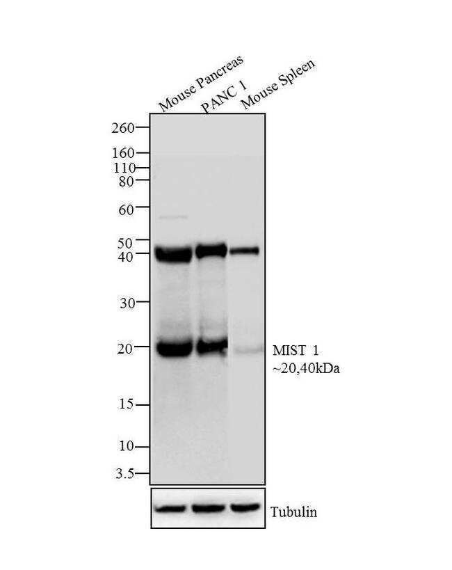
- Western blot analysis was performed on whole cell extracts (30 µg lysate) of Mouse Pancreas (Lane 1), Panc1 (Lane 2) and Mouse spleen (Lane 3). The blots were probed with Anti-Mist 1 Mouse Monoclonal Antibody (Product # MA1-517, 1:100-1:500 dilution) and detected by chemiluminescence Goat anti-Mouse IgG (H+L) Secondary Antibody, HRP conjugate (Product # 62-6520, 1:4000 dilution). Expected molecular weight is 20 kDa, a 40 kDa band was observed across cell lines tested which may be due to acetylation of Mist1 protein. Known quantity of protein samples were electrophoresed using Novex® NuPAGE® 4-12 % Bis-Tris gel (Product # NP0321BOX), XCell SureLock™ Electrophoresis System (Product # EI0002) and Novex® Sharp Pre-Stained Protein Standard (Product # LC5800). Resolved proteins were then transferred onto a nitrocellulose membrane by Pierce™ Power Blotter System (22834).The membrane was probed with the relevant primary and secondary Antibody following blocking with 5 % skimmed milk. Chemiluminescent detection was performed using Pierce™ ECL Western Blotting Substrate (Product # 32106).
Supportive validation
- Submitted by
- Invitrogen Antibodies (provider)
- Main image

- Experimental details
- Immunohistochemical staining of paraffin embedded mouse pancreas tissues using Product # MA1-517. The antibody gives nuclear staining in acinar cells but not in duct or islet cells.

- Submitted by
- Invitrogen Antibodies (provider)
- Main image

- Experimental details
- Immunohistochemical staining of paraffin embedded mouse pancreas tissues using Product # MA1-517. The antibody gives nuclear staining in acinar cells but not in duct or islet cells.

Supportive validation
- Submitted by
- Invitrogen Antibodies (provider)
- Main image

- Experimental details
- Figure 1 Expression of ACE2 and TMPRSS2 in exocrine pancreatic cells. (A) iPan EXO Ductal cells CK19 (red) exhibiting i . ACE2 (green) and ii. TMPRSS2 (green) expression. (B) iPan EXO Acinar cells Amylase (red), MIST1 (gray) exhibit i . ACE2 (green) expression and ii. iPan EXO Acinar CTRC (red) exhibit TMPRSS2 (green). (C) iPan EXO Acinar and iPan ENDO cultures contain some endocrine C-peptide expressing cells, which also co-stain with ACE2. (D) iPSC-derived pancreatic exocrine cells as well as human acinar tissues and human ductal cell line H6C7 express ACE2 and TMPRSS2 . Data is shown as mean +- SEM with statistical significance determined by unpaired two-tailed t-test. **p < 0.01 and ****p < 0.0001. Scale bar represents 100 um, and 20 um for zoomed panels adjacent to main images. ICC images shown here are representative results from 27 independent sites acquired. RNA was extracted from aggregates of 2-3 biological replicates (using 12 well-plates), and qPCR was run with 3 technical replicates per sample. These results were pooled from 2 independent rounds of infection experiments.
