Explore
 Validate
 Learn
Learn Western blot
Antibody data
- Antibody Data
- Antigen structure
- References [1]
- Comments [0]
- Validations
- Western blot [2]
- Immunocytochemistry [1]
Submit
Validation data
Reference
Comment
Report error
- Product number
- PA5-26336 - Provider product page

- Provider
- Invitrogen Antibodies
- Product name
- DLL3 Polyclonal Antibody
- Antibody type
- Polyclonal
- Antigen
- Synthetic peptide
- Reactivity
- Human, Mouse, Rat
- Host
- Rabbit
- Vial size
- 200 µL
- Concentration
- 0.5 mg/mL
- Storage
- -20° C, Avoid Freeze/Thaw Cycles
Submitted references Comparison of four DLL3 antibodies performance in high grade neuroendocrine lung tumor samples and cell cultures.
Brcic L, Kuchler C, Eidenhammer S, Pabst D, Quehenberger F, Gazdar AF, Popper H
Diagnostic pathology 2019 May 20;14(1):47
Diagnostic pathology 2019 May 20;14(1):47
No comments: Submit comment
Supportive validation
- Submitted by
- Invitrogen Antibodies (provider)
- Main image

- Experimental details
- Western blot analysis using a DLL3 polyclonal antibody (Product # PA5-26336) in A375 cell lysates (35 µg per lane).

- Submitted by
- Invitrogen Antibodies (provider)
- Main image

- Experimental details
- Western blot analysis was performed on whole cell extracts of U-87 MG (Lane 1), SH-SY5Y (Lane 2), A549 (Lane 3), A-375 (Lane 4), HeLa (Lane 5), HEK-293 (Lane 6), tissue extracts of Mouse Pup Brain (Lane 7), Mouse Brain (Lane 8) and Rat Brain (Lane 9). The blot was probed with Anti-DLL3 Polyclonal Antibody (Product # PA5-26336, 1:2000 dilution) and detected by chemiluminescence using Goat anti-Rabbit IgG (H+L) Superclonal™ Secondary Antibody, HRP conjugate (Product # A27036, 0.25 µg/ml, 1:4000 dilution). A 62 kDa band corresponding to DLL3 was observed across the cell lines and tissues tested.

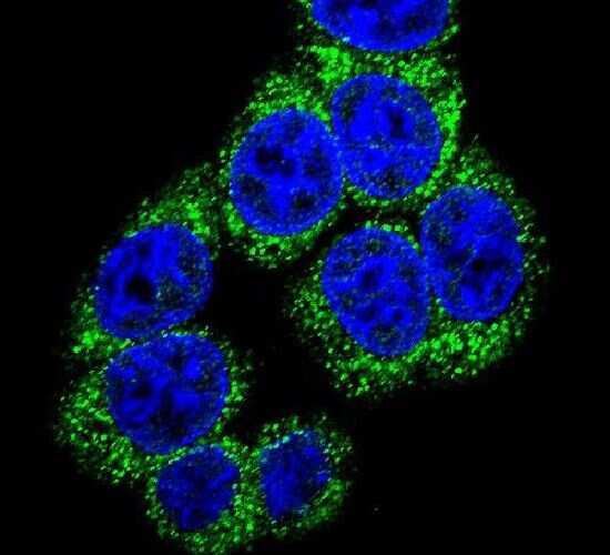
Supportive validation
- Submitted by
- Invitrogen Antibodies (provider)
- Main image
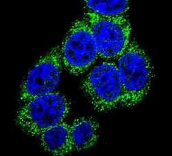
- Experimental details
- Immunofluorescent analysis of 293 cells using a DLL3 polyclonal antibody (Product # PA5-26336) at a dilution of 1:10-50, followed by a fluor-conjugated goat anti-rabbit secondary antibody (green). Nuclei were stained with DAPI (blue).