Explore
 Validate
 Learn
Learn Western blot
Antibody data
- Antibody Data
- Antigen structure
- References [0]
- Comments [0]
- Validations
- Western blot [1]
- Immunocytochemistry [1]
- Flow cytometry [1]
Submit
Validation data
Reference
Comment
Report error
- Product number
- MAB5447 - Provider product page

- Provider
- R&D Systems
- Product name
- Human Galectin-10 Antibody
- Antibody type
- Monoclonal
- Description
- Protein A or G purified from hybridoma culture supernatant. Detects human Galectin-10 in direct ELISAs and Western blots. In direct ELISAs, no cross-reactivity with recombinant human Galectin-1, -2, -3, -4, -7, -8, or -9/Ecalectin is observed.
- Reactivity
- Human
- Host
- Mouse
- Conjugate
- Unconjugated
- Antigen sequence
Q05315- Isotype
- IgG
- Antibody clone number
- 561603
- Vial size
- 100 ug
- Concentration
- LYOPH
- Storage
- Use a manual defrost freezer and avoid repeated freeze-thaw cycles. 12 months from date of receipt, -20 to -70 °C as supplied. 1 month, 2 to 8 °C under sterile conditions after reconstitution. 6 months, -20 to -70 °C under sterile conditions after reconstitution.
No comments: Submit comment
Supportive validation
- Submitted by
- R&D Systems (provider)
- Main image
- Experimental details
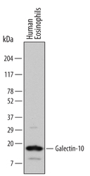
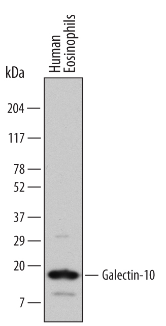
- Detection of Human Galectin-10 by Western Blot. Western blot shows lysates of human eosinophils (enriched, approximately 60%). PVDF Membrane was probed with 1 µg/mL of Human Galectin-10 Monoclonal Antibody (Catalog # MAB5447) followed by HRP-conjugated Anti-Mouse IgG Secondary Antibody (Catalog # HAF007). A specific band was detected for Galectin-10 at approximately 16 kDa (as indicated). This experiment was conducted under reducing conditions and using Immunoblot Buffer Group 1.
Supportive validation
- Submitted by
- R&D Systems (provider)
- Main image
- Experimental details
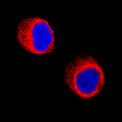
- Galectin-10 in HL-60 Human Cell Line. Galectin-10 was detected in immersion fixed HL-60 human acute promyelocytic leukemia cell line using Mouse Anti-Human Galectin-10 Monoclonal Antibody (Catalog # MAB5447) at 8 µg/mL for 3 hours at room temperature. Cells were stained using the NorthernLights™ 557-conjugated Anti-Mouse IgG Secondary Antibody (red; Catalog # NL007) and counterstained with DAPI (blue). Specific staining was localized to cytoplasm. View our protocol for Fluorescent ICC Staining of Non-adherent Cells.

Supportive validation
- Submitted by
- R&D Systems (provider)
- Main image

- Experimental details
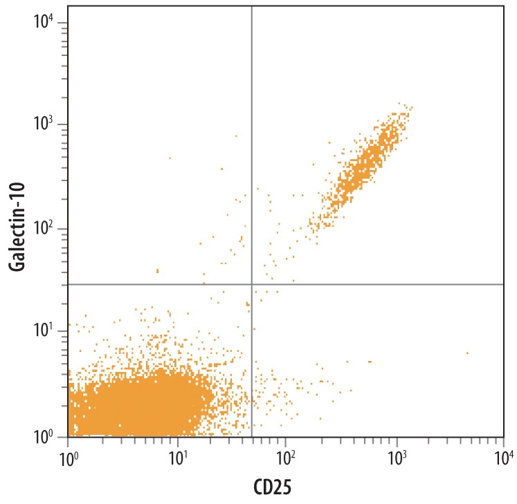
- Detection of Galectin-10 in Human PBMCs by Flow Cytometry. Human peripheral blood lymphocytes were stained with Human Galectin-10 Monoclonal Antibody (Catalog # MAB5447) followed by Fluorescein-conjugated Anti-Mouse IgG Secondary Antibody (Catalog # F0103B) and Human IL-2 R alpha APC-conjugated Monoclonal Antibody (Catalog # FAB1020A). Quadrant markers were set based on control antibody staining (Catalog # IC0041F and IC003A, respectively).