 Explore
 Validate
 Learn
Learn Western blot
 ELISA
ELISAAntibody data
- Antibody Data
- Antigen structure
- References [0]
- Comments [0]
- Validations
- Western blot [8]
- ELISA [2]
Submit
Validation data
Reference
Comment
Report error
- Product number
- LS-C829082 - Provider product page

- Provider
- LSBio
- Product name
- WDTC1 Antibody (clone 8C8) LS-C829082
- Antibody type
- Monoclonal
- Description
- Protein A purified
- Reactivity
- Mouse
- Host
- Mouse
- Isotype
- IgG
- Antibody clone number
- 8C8
- Storage
- Store lyophilized at -20°C for up to 1 year. Store reconstituted at 2°C to 8°C for up to 3 weeks. Aliquot and store at -20°C or below for up to 1 year. Avoid freeze/thaw cycles.
No comments: Submit comment
Supportive validation
- Submitted by
- LSBio (provider)
- Main image

- Experimental details
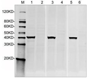
- Western Blot of Mouse Wdtc1 recombinant protein with two independent antibodies: Mouse Wdtc1 Antibody (8C8) and Mouse Wdtc1 Antibody (1B12). The correlated pattern indicates the high specificity of these two antibodies. Lane 1: 50 ng Mouse Wdtc1 recombinant protein. Lane 2: 25 ng Mouse Wdtc1 recombinant protein. Lane 3: 10 ng Mouse Wdtc1 recombinant protein. Lane 4: 50 ng Mouse Wdtc1 recombinant protein. Lane 5: 25 ng Mouse Wdtc1 recombinant protein. Lane 6: 10 ng Mouse Wdtc1 recombinant protein. Primary Antibody: Lane 1~3: Mouse Wdtc1 Antibody (1B12) 1 µg/ml. Lane 4~6: Mouse Wdtc1 Antibody (8C8) 1 µg/ml. Secondary Antibody: Goat anti-Mouse IgG (H&L) [IRDye800] 0.125 µg/ml.

- Submitted by
- LSBio (provider)
- Main image

- Experimental details
- Western Blot of Wild-type and Wdtc1 knockout HepG2 cell lysates with Mouse Wdtc1 Antibody (8C8). The different concentration of antibodies indicates the high specificity and sensitivity of the antibody. Lane 1: 50 µg Wild-type HepG2 cell Lysate. Lane 2: 50 µg Wdtc1 knockout HepG2 cell Lysate. Lane 3: 50 µg Wild-type HepG2 cell Lysate. Lane 4: 50 µg Wdtc1 knockout HepG2 cell Lysate. Lane 5: 50 µg Wild-type HepG2 cell Lysate. Lane 6: 50 µg Wdtc1 knockout HepG2 cell Lysate. Primary Antibody: Lane 1~2: Mouse Wdtc1 Antibody (8C8) 2.5 µg/ml. Lane 3~4: Mouse Wdtc1 Antibody (8C8) 1 µg/ml. Lane 5~6: Mouse Wdtc1 Antibody (8C8) 0.5 µg/ml. Secondary Antibody: Goat anti-Mouse IgG (H&L) [IRDye800] 0.125 µg/ml.
- Submitted by
- LSBio (provider)
- Main image
- Experimental details
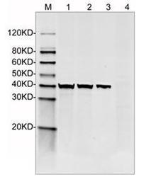
- Western Blot of Wild-type and Wdtc1 knockout HepG2 cell lysates with Mouse Wdtc1 Antibody (8C8). The different concentration of cell lysates indicates the high specificity and sensitivity of the antibody. Lane 1: 50 µg Wild-type HepG2 cell Lysate. Lane 2: 25 µg Wild-type HepG2 cell Lysate. Lane 3: 10 µg Wild-type HepG2 cell Lysate. Lane 4: 50 µg Wdtc1 knockout HepG2 cell Lysate. Primary Antibody: Mouse Wdtc1 Antibody (8C8) 1 µg/ml. Secondary Antibody: Goat anti-Mouse IgG (H&L) [IRDye800] 0.125 µg/ml.

- Submitted by
- LSBio (provider)
- Main image
- Experimental details
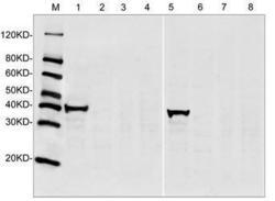
- Western Blot of Wild-type and Wdtc1 knockout HepG2 cell lysates with two independent antibodies: Mouse Wdtc1 Antibody (8C8) and Mouse Wdtc1 Antibody (1B12). The correlated pattern minimizes the likelihood of off-target binding to unrelated protein. Lane 1: 50 µg Wild-type HepG2 cell Lysate. Lane 2: 50 µg Wdtc1 knockout HepG2 cell Lysate. Lane 3: 50 µg Wdtc1 knockout HepG2 cell Lysate. Lane 4: 50 µg Wdtc1 knockout HepG2 cell Lysate. Lane 5: 50 µg Wild-type HepG2 cell Lysate. Lane 6: 50 µg Wdtc1 knockout HepG2 cell Lysate. Lane 7: 50 µg Wdtc1 knockout HepG2 cell Lysate. Lane 8: 50 µg Wdtc1 knockout HepG2 cell Lysate. Primary Antibody: Lane 1~4: Mouse Wdtc1 Antibody (1B12) 1 µg/ml. Lane 5~8: Mouse Wdtc1 Antibody (8C8) 1 µg/ml. Secondary Antibody: Goat anti-Mouse IgG (H&L) [IRDye800] 0.125 µg/ml.

- Submitted by
- LSBio (provider)
- Main image

- Experimental details
- Western Blot of Mouse Wdtc1 recombinant protein with two independent antibodies: Mouse Wdtc1 Antibody (8C8) and Mouse Wdtc1 Antibody (1B12). The correlated pattern indicates the high specificity of these two antibodies. Lane 1: 50 ng Mouse Wdtc1 recombinant protein. Lane 2: 25 ng Mouse Wdtc1 recombinant protein. Lane 3: 10 ng Mouse Wdtc1 recombinant protein. Lane 4: 50 ng Mouse Wdtc1 recombinant protein. Lane 5: 25 ng Mouse Wdtc1 recombinant protein. Lane 6: 10 ng Mouse Wdtc1 recombinant protein. Primary Antibody: Lane 1~3: Mouse Wdtc1 Antibody (1B12) 1 µg/ml. Lane 4~6: Mouse Wdtc1 Antibody (8C8) 1 µg/ml. Secondary Antibody: Goat anti-Mouse IgG (H&L) [IRDye800] 0.125 µg/ml.

- Submitted by
- LSBio (provider)
- Main image

- Experimental details
- Western Blot of Wild-type and Wdtc1 knockout HepG2 cell lysates with Mouse Wdtc1 Antibody (8C8). The different concentration of antibodies indicates the high specificity and sensitivity of the antibody. Lane 1: 50 µg Wild-type HepG2 cell Lysate. Lane 2: 50 µg Wdtc1 knockout HepG2 cell Lysate. Lane 3: 50 µg Wild-type HepG2 cell Lysate. Lane 4: 50 µg Wdtc1 knockout HepG2 cell Lysate. Lane 5: 50 µg Wild-type HepG2 cell Lysate. Lane 6: 50 µg Wdtc1 knockout HepG2 cell Lysate. Primary Antibody: Lane 1~2: Mouse Wdtc1 Antibody (8C8) 2.5 µg/ml. Lane 3~4: Mouse Wdtc1 Antibody (8C8) 1 µg/ml. Lane 5~6: Mouse Wdtc1 Antibody (8C8) 0.5 µg/ml. Secondary Antibody: Goat anti-Mouse IgG (H&L) [IRDye800] 0.125 µg/ml.

- Submitted by
- LSBio (provider)
- Main image

- Experimental details
- Western Blot of Wild-type and Wdtc1 knockout HepG2 cell lysates with Mouse Wdtc1 Antibody (8C8). The different concentration of cell lysates indicates the high specificity and sensitivity of the antibody. Lane 1: 50 µg Wild-type HepG2 cell Lysate. Lane 2: 25 µg Wild-type HepG2 cell Lysate. Lane 3: 10 µg Wild-type HepG2 cell Lysate. Lane 4: 50 µg Wdtc1 knockout HepG2 cell Lysate. Primary Antibody: Mouse Wdtc1 Antibody (8C8) 1 µg/ml. Secondary Antibody: Goat anti-Mouse IgG (H&L) [IRDye800] 0.125 µg/ml.

- Submitted by
- LSBio (provider)
- Main image

- Experimental details
- Western Blot of Wild-type and Wdtc1 knockout HepG2 cell lysates with two independent antibodies: Mouse Wdtc1 Antibody (8C8) and Mouse Wdtc1 Antibody (1B12). The correlated pattern minimizes the likelihood of off-target binding to unrelated protein. Lane 1: 50 µg Wild-type HepG2 cell Lysate. Lane 2: 50 µg Wdtc1 knockout HepG2 cell Lysate. Lane 3: 50 µg Wdtc1 knockout HepG2 cell Lysate. Lane 4: 50 µg Wdtc1 knockout HepG2 cell Lysate. Lane 5: 50 µg Wild-type HepG2 cell Lysate. Lane 6: 50 µg Wdtc1 knockout HepG2 cell Lysate. Lane 7: 50 µg Wdtc1 knockout HepG2 cell Lysate. Lane 8: 50 µg Wdtc1 knockout HepG2 cell Lysate. Primary Antibody: Lane 1~4: Mouse Wdtc1 Antibody (1B12) 1 µg/ml. Lane 5~8: Mouse Wdtc1 Antibody (8C8) 1 µg/ml. Secondary Antibody: Goat anti-Mouse IgG (H&L) [IRDye800] 0.125 µg/ml.

Enhanced validation
- Submitted by
- LSBio (provider)
- Enhanced method
- Genetic validation
- Main image

- Experimental details
- Standard curve of Wdtc1 Sandwich ELISA. The Wdtc1 Sandwich ELISA assay is developed by using Mouse Wdtc1 Antibody (1B12) and Mouse Wdtc1 Antibody (8C8) as capture and detection antibody, respectively. These two antibodies recognize different epitopes. In this ELISA assay, Mouse Wdtc1 Antibody (8C8) was labeled with Biotin. The sensitivity is <30 ng/ml and the detection range is 0-1000 ng/ml.

- Submitted by
- LSBio (provider)
- Main image

- Experimental details
- Standard curve of Wdtc1 Sandwich ELISA. The Wdtc1 Sandwich ELISA assay is developed by using Mouse Wdtc1 Antibody (1B12) and Mouse Wdtc1 Antibody (8C8) as capture and detection antibody, respectively. These two antibodies recognize different epitopes. In this ELISA assay, Mouse Wdtc1 Antibody (8C8) was labeled with Biotin. The sensitivity is <30 ng/ml and the detection range is 0-1000 ng/ml.
