 Explore
 Validate
 Learn
LearnMAB4675
antibody from R&D Systems
Targeting: CRISP1
AEGL1, ARP, CRISP-1, HSCRISP1D, HSCRISP1G, HUMARP
 Western blot
Antibody data
- Antibody Data
- Antigen structure
- References [0]
- Comments [0]
- Validations
- Western blot [1]
- Immunohistochemistry [1]
- Blocking/Neutralizing [1]
Submit
Validation data
Reference
Comment
Report error
- Product number
- MAB4675 - Provider product page

- Provider
- R&D Systems
- Product name
- Mouse CRISP-1 Antibody
- Antibody type
- Monoclonal
- Description
- Protein A or G purified from hybridoma culture supernatant. Detects mouse CRISP-1 in direct ELISAs.
- Reactivity
- Mouse
- Host
- Rat
- Conjugate
- Unconjugated
- Antigen sequence
Q03401- Isotype
- IgG
- Antibody clone number
- 840017
- Vial size
- 100 ug
- Storage
- Use a manual defrost freezer and avoid repeated freeze-thaw cycles. 12 months from date of receipt, -20 to -70 °C as supplied. 1 month, 2 to 8 °C under sterile conditions after reconstitution. 6 months, -20 to -70 °C under sterile conditions after reconstitution.
No comments: Submit comment
Supportive validation
- Submitted by
- R&D Systems (provider)
- Main image
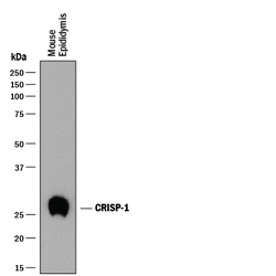
- Experimental details
- Detection of Mouse CRISP-1 by Western Blot. Western blot shows lysates of mouse epididymis tissue. PVDF membrane was probed with 1 µg/mL of Rat Anti-Mouse CRISP-1 Monoclonal Antibody (Catalog # MAB4675) followed by HRP-conjugated Anti-Rat IgG Secondary Antibody (Catalog # HAF005). A specific band was detected for CRISP-1 at approximately 27-30 kDa (as indicated). This experiment was conducted under reducing conditions and using Immunoblot Buffer Group 1.

Supportive validation
- Submitted by
- R&D Systems (provider)
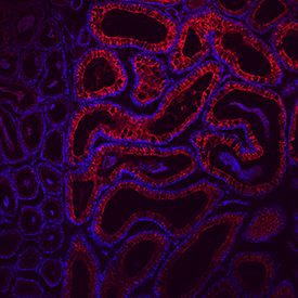
- Main image
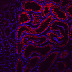
- Experimental details
- CRISP-1 in Mouse Epididymis. CRISP-1 was detected in perfusion fixed frozen sections of mouse epididymis using Rat Anti-Mouse CRISP-1 Monoclonal Antibody (Catalog # MAB4675) at 10 µg/mL overnight at 4 °C. Tissue was stained using the NorthernLights™ 557-conjugated Anti-Rat IgG Secondary Antibody (red; Catalog # NL013) and counterstained with DAPI (blue). Specific staining was localized to cytoplasm. View our protocol for Fluorescent IHC Staining of Frozen Tissue Sections.
Supportive validation
- Submitted by
- R&D Systems (provider)
- Main image

- Experimental details
- Proliferation Inhibited by CRISP-1 and Neutralization by Mouse CRISP-1 Antibody. Recombinant Mouse CRISP-1 inhibits proliferation in the 3A-sub E human placenta cell line in a dose-dependent manner (orange line), as measured by Resazurin (Catalog # AR002). Proliferation inhibited by Recombinant Mouse CRISP-1 (10 µg/mL) is neutralized (green line) by increasing concentrations of Rat Anti-Mouse CRISP-1 Monoclonal Antibody (Catalog # MAB4675). The ND50 is typically 10-20 µg/mL.
