Explore
 Validate
 Learn
Learn Western blot
 ELISA
ELISAAntibody data
- Antibody Data
- Antigen structure
- References [0]
- Comments [0]
- Validations
- Western blot [2]
Submit
Validation data
Reference
Comment
Report error
- Product number
- AP09245PU-N - Provider product page

- Provider
- Acris Antibodies GmbH
- Proper citation
- Acris Antibodies GmbH Cat#AP09245PU-N, RRID:AB_2035463
- Product name
- anti FANCA (995-1009)
- Antibody type
- Polyclonal
- Antigen
- Synthetic peptide corresponding to amino acids 995-1009 of human FANCA protein
- Reactivity
- Human
- Host
- Rabbit
- Isotype
- IgG
- Vial size
- 0.1 mg
- Concentration
- 1.45 mg/ml (by UV absorbance at 280 nm)
No comments: Submit comment
Supportive validation
- Submitted by
- Acris Antibodies GmbH (provider)
- Main image
- Experimental details
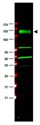
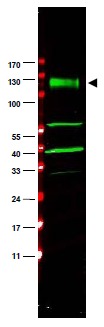
- Western blot using anti-FANCA antibody shows detection of a band at ~133 kDa (arrowhead) corresponding to FANCA in HeLa whole cell lysates. The identity of the lower molecular weight bands is unknown but may represent breakdown products. Approximately 35 μg of lysate was separated by 4-20% Tris Glycine SDS-PAGE. After blocking, the membrane was probed for 2 h at room temperature with the primary antibody diluted to 1:1,500. The membrane was washed and reacted with a 1:10,000 dilution of IRDye(TM)800 conjugated Gt-a-Rabbit IgG [H&L] for 45 min at room temperature (800 nm channel, green). Molecular weight estimation was made by comparison to prestained MW markers indicated at left (700 nm channel, red). IRDye(TM)800 fluorescence images were captured using the Odyssey® Infrared Imaging System developed by LI-COR. IRDye is a trademark of LI-COR, Inc. Other detection systems will yield similar results.
- Submitted by
- Acris Antibodies GmbH (provider)
- Main image
- Experimental details
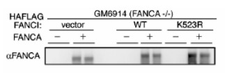
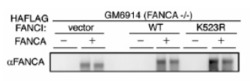
- Western blot using anti-FANCA antibody shows detection of FANCA only in FANCA transfected GM6914 cell lysates. No staining is seen in lysates prepared from FANCA (-/-) cells in the absence of FANCA transfection. Modified from Smogorzewska et al (2007) Cell 129, 289-301.