 Explore
 Validate
 Learn
Learn Western blot
Antibody data
- Antibody Data
- Antigen structure
- References [1]
- Comments [0]
- Validations
- Western blot [1]
- Immunohistochemistry [14]
- Other assay [1]
Submit
Validation data
Reference
Comment
Report error
- Product number
- PA5-115697 - Provider product page

- Provider
- Invitrogen Antibodies
- Product name
- SP7 Polyclonal Antibody
- Antibody type
- Polyclonal
- Antigen
- Synthetic peptide
- Reactivity
- Human, Mouse, Rat
- Host
- Rabbit
- Isotype
- IgG
- Vial size
- 100 µL
- Concentration
- 1 mg/mL
- Storage
- -20°C
Submitted references In vitro evaluation of periapical lesion-derived stem cells for dental pulp tissue engineering.
Li W, Mao M, Hu N, Wang J, Huang J, Gu S
FEBS open bio 2022 Jan;12(1):270-284
FEBS open bio 2022 Jan;12(1):270-284
No comments: Submit comment
Supportive validation
- Submitted by
- Invitrogen Antibodies (provider)
- Main image

- Experimental details
- Western blot analysis of SP7 in K562 whole lysates. Samples were incubated with polyclonal antibody (Product # PA5-115697).

Supportive validation
- Submitted by
- Invitrogen Antibodies (provider)
- Main image
- Experimental details
- Immunohistochemistry analysis of SP7 in rat thymus tissue. Samples were treated with formaldehyde and treated with citrate buffer for antigen retrieval, blocked, and incubated (1.5 hours at 22°C) with polyclonal antibody (Product # PA5-115697) at a dilution of 1:100. Secondary staining was applied with HRP conjugated anti-Rabbit.

- Submitted by
- Invitrogen Antibodies (provider)
- Main image

- Experimental details
- Immunohistochemistry analysis of SP7 in rat lung tissue. Samples were treated with formaldehyde and treated with citrate buffer for antigen retrieval, blocked, and incubated (1.5 hours at 22°C) with polyclonal antibody (Product # PA5-115697) at a dilution of 1:100. Secondary staining was applied with HRP conjugated anti-Rabbit.

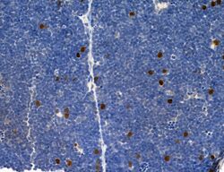
- Submitted by
- Invitrogen Antibodies (provider)
- Main image

- Experimental details
- Immunohistochemistry analysis of SP7 in human normal tissues adjacent to mammary cancer. Samples were treated with formaldehyde and treated with citrate buffer for antigen retrieval, blocked, and incubated (1.5 hours at 22°C) with polyclonal antibody (Product # PA5-115697) at a dilution of 1:100. Secondary staining was applied with HRP conjugated anti-Rabbit.

- Submitted by
- Invitrogen Antibodies (provider)
- Main image

- Experimental details
- Immunohistochemistry analysis of SP7 in rat ovary tissue. Samples were treated with formaldehyde and treated with citrate buffer for antigen retrieval, blocked, and incubated (1.5 hours at 22°C) with polyclonal antibody (Product # PA5-115697) at a dilution of 1:100. Secondary staining was applied with HRP conjugated anti-Rabbit.
- Submitted by
- Invitrogen Antibodies (provider)
- Main image
- Experimental details
- Immunohistochemistry analysis of SP7 in rat stomach tissue. Samples were treated with formaldehyde and treated with citrate buffer for antigen retrieval, blocked, and incubated (1.5 hours at 22°C) with polyclonal antibody (Product # PA5-115697) at a dilution of 1:100. Secondary staining was applied with HRP conjugated anti-Rabbit.

- Submitted by
- Invitrogen Antibodies (provider)
- Main image

- Experimental details
- Immunohistochemistry analysis of SP7 in rat testis tissue. Samples were treated with formaldehyde and treated with citrate buffer for antigen retrieval, blocked, and incubated (1.5 hours at 22°C) with polyclonal antibody (Product # PA5-115697) at a dilution of 1:100. Secondary staining was applied with HRP conjugated anti-Rabbit.

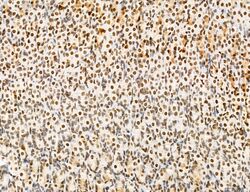
- Submitted by
- Invitrogen Antibodies (provider)
- Main image

- Experimental details
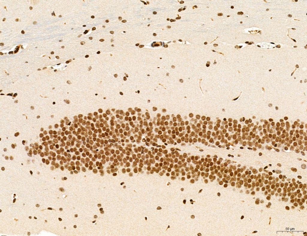
- Immunohistochemistry analysis of SP7 in rat brain tissue. Samples were treated with formaldehyde and treated with citrate buffer for antigen retrieval, blocked, and incubated (1.5 hours at 22°C) with polyclonal antibody (Product # PA5-115697) at a dilution of 1:100. Secondary staining was applied with HRP conjugated anti-Rabbit.

- Submitted by
- Invitrogen Antibodies (provider)
- Main image
- Experimental details
- Immunohistochemistry analysis of SP7 in mouse stomach tissue. Samples were treated with formaldehyde and treated with citrate buffer for antigen retrieval, blocked, and incubated (1.5 hours at 22°C) with polyclonal antibody (Product # PA5-115697) at a dilution of 1:100. Secondary staining was applied with HRP conjugated anti-Rabbit.

- Submitted by
- Invitrogen Antibodies (provider)
- Main image

- Experimental details
- Immunohistochemistry analysis of SP7 in mouse testis tissue. Samples were treated with formaldehyde and treated with citrate buffer for antigen retrieval, blocked, and incubated (1.5 hours at 22°C) with polyclonal antibody (Product # PA5-115697) at a dilution of 1:100. Secondary staining was applied with HRP conjugated anti-Rabbit.

- Submitted by
- Invitrogen Antibodies (provider)
- Main image

- Experimental details
- Immunohistochemistry analysis of SP7 in mouse colon tissue. Samples were treated with formaldehyde and treated with citrate buffer for antigen retrieval, blocked, and incubated (1.5 hours at 22°C) with polyclonal antibody (Product # PA5-115697) at a dilution of 1:100. Secondary staining was applied with HRP conjugated anti-Rabbit.

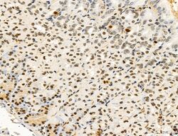
- Submitted by
- Invitrogen Antibodies (provider)
- Main image

- Experimental details
- Immunohistochemistry analysis of SP7 in mouse brain tissue. Samples were treated with formaldehyde and treated with citrate buffer for antigen retrieval, blocked, and incubated (1.5 hours at 22°C) with polyclonal antibody (Product # PA5-115697) at a dilution of 1:100. Secondary staining was applied with HRP conjugated anti-Rabbit.
- Submitted by
- Invitrogen Antibodies (provider)
- Main image

- Experimental details
- Immunohistochemistry analysis of SP7 in mouse skin tissue. Samples were treated with formaldehyde and treated with citrate buffer for antigen retrieval, blocked, and incubated (1.5 hours at 22°C) with polyclonal antibody (Product # PA5-115697) at a dilution of 1:100. Secondary staining was applied with HRP conjugated anti-Rabbit.

- Submitted by
- Invitrogen Antibodies (provider)
- Main image

- Experimental details
- Immunohistochemistry analysis of SP7 in rat spleen tissue. Samples were treated with formaldehyde and treated with citrate buffer for antigen retrieval, blocked, and incubated (1.5 hours at 22°C) with polyclonal antibody (Product # PA5-115697) at a dilution of 1:100. Secondary staining was applied with HRP conjugated anti-Rabbit.

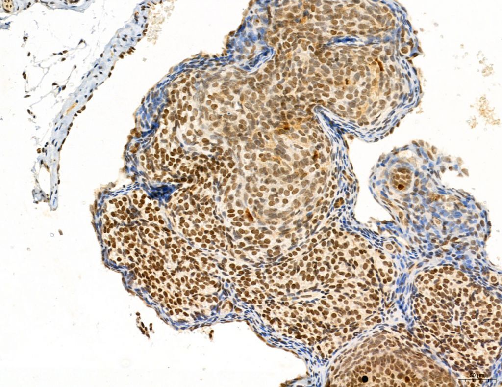
- Submitted by
- Invitrogen Antibodies (provider)
- Main image

- Experimental details
- Immunohistochemistry analysis of SP7 in human normal tissues adjacent to colorectal cancer. Samples were treated with formaldehyde and treated with citrate buffer for antigen retrieval, blocked, and incubated (1.5 hours at 22°C) with polyclonal antibody (Product # PA5-115697) at a dilution of 1:100. Secondary staining was applied with HRP conjugated anti-Rabbit.

Supportive validation
- Submitted by
- Invitrogen Antibodies (provider)
- Main image

- Experimental details
- Fig. 3 Comparison of the osteo/odontogenic capacities of the cells. (A) ALP staining after osteo/odontogenic induction for 3 and 7 days. Scale bar = 500 mum. (B) Alizarin red staining showing the presence of calcified nodules after 7, 14, 21 and 28 days of osteo/odontogenic induction. Scale bar = 500 mum. (C) Gene expression patterns of ALP , RUNX2 and DSPP after osteogenic induction for 3 and 7 days; the expression level was normalized to that of ACTB ( n = 3, Student's t -test). * P < 0.05, ** P < 0.01. (D) Western blotting detection (upper) and imagej analysis (below) of DSPP, DMP1, RUNX2 and SP7 after osteogenic induction for 7, 14 and 21 days. Data are shown as the mean +- SD.
