Explore
 Validate
 Learn
Learn Immunocytochemistry
Antibody data
- Antibody Data
- Antigen structure
- References [0]
- Comments [0]
- Validations
- Immunocytochemistry [2]
- Immunohistochemistry [3]
Submit
Validation data
Reference
Comment
Report error
- Product number
- PA1-1755 - Provider product page

- Provider
- Invitrogen Antibodies
- Product name
- ITIH1 Polyclonal Antibody
- Antibody type
- Polyclonal
- Antigen
- Synthetic peptide
- Description
- PA1-1755 detects HC1 from human and mouse samples.
- Concentration
- 1 mg/mL
No comments: Submit comment
Supportive validation
- Submitted by
- Invitrogen Antibodies (provider)
- Main image
- Experimental details
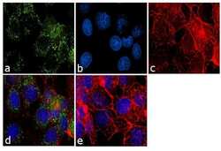
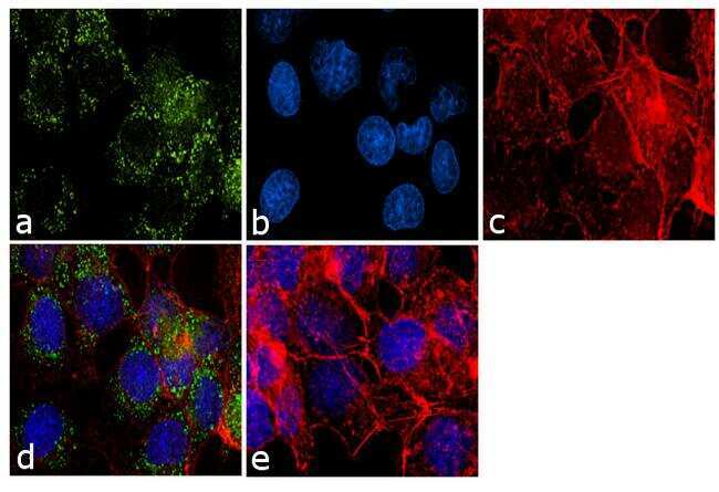
- Immunofluorescence analysis of ITIH1 was performed using 70% confluent log phase HepG2 cells. The cells were fixed with 4% paraformaldehyde for 10 minutes, permeabilized with 0.1% Triton™ X-100 for 10 minutes, and blocked with 1% BSA for 1 hour at room temperature. The cells were labeled with ITIH1 Rabbit Polyclonal Antibody (Product # PA1-1755) at 2µg/mLin 0.1% BSA and incubated for 3 hours at room temperature and then labeled with Goat anti-Rabbit IgG (H+L) Superclonal™ Secondary Antibody, Alexa Fluor® 488 conjugate (Product # A27034) at a dilution of 1:2000 for 45 minutes at room temperature (Panel a: green). Nuclei (Panel b: blue) were stained with SlowFade® Gold Antifade Mountant with DAPI (Product # S36938). F-actin (Panel c: red) was stained with Rhodamine Phalloidin (Product # R415, 1:300). Panel d represents the merged image showing cytoplasmic localization. Panel e shows the no primary antibody control. The images were captured at 60X magnification.
- Submitted by
- Invitrogen Antibodies (provider)
- Main image
- Experimental details
- Immunofluorescent analysis of ITIH1 using a polyclonal antibody (Product # PA1-1755).

Supportive validation
- Submitted by
- Invitrogen Antibodies (provider)
- Main image

- Experimental details
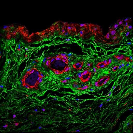
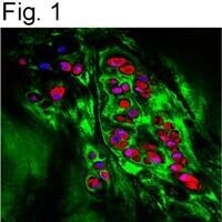
- Figure 1 illustrates immunostaining of HC1 on paraffin embedded human OA chondrocyte tissue using Product # PA1-1755. HC1 is stained in red, hyaluronan in green, and the nuclei in blue.
- Submitted by
- Invitrogen Antibodies (provider)
- Main image
- Experimental details
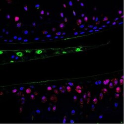
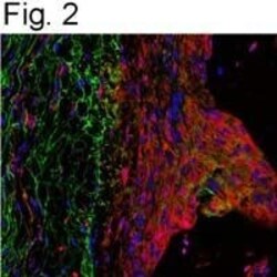
- Figure 2 illustrates immunostaining of HC1 on paraffin embedded mouse OA periosteum tissue using Product # PA1-1755. HC1 is stained in red, hyaluronan in green, and the nuclei in blue.

- Submitted by
- Invitrogen Antibodies (provider)
- Main image
- Experimental details
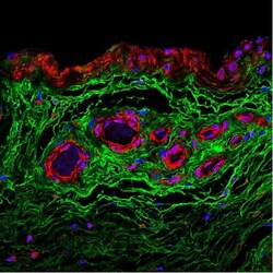
- Immunostaining of HC1 on paraffin embedded human OA chondrocyte tissue using Product # PA1-1755. HC1 is stained in red, hyaluronan in green, and the nuclei in blue.