 Explore
 Validate
 Learn
Learn Western blot
 Immunohistochemistry
Antibody data
- Antibody Data
- Antigen structure
- References [3]
- Comments [0]
- Validations
- Western blot [1]
- Immunohistochemistry [8]
Submit
Validation data
Reference
Comment
Report error
- Product number
- HPA031700 - Provider product page

- Provider
- Atlas Antibodies
- Proper citation
- Atlas Antibodies Cat#HPA031700, RRID:AB_10601693
- Product name
- Anti-MB21D1
- Antibody type
- Polyclonal
- Reactivity
- Human
- Host
- Rabbit
- Conjugate
- Unconjugated
- Antigen sequence
RKQLRLKPFYLVPKHAKEGNGFQEETWRLSFSHIE
KEILNNHGKSKTCCENKEEKCCRKDCLKLMKYLLE
QLKERF- Isotype
- IgG
- Vial size
- 100 µl
- Storage
- Store at +4°C for short term storage. Long time storage is recommended at -20°C.
Submitted references T cells detect intracellular DNA but fail to induce type I IFN responses: implications for restriction of HIV replication.
Pan-viral specificity of IFN-induced genes reveals new roles for cGAS in innate immunity.
Listeria monocytogenes induces IFNβ expression through an IFI16-, cGAS- and STING-dependent pathway.
Berg RK, Rahbek SH, Kofod-Olsen E, Holm CK, Melchjorsen J, Jensen DG, Hansen AL, Jørgensen LB, Ostergaard L, Tolstrup M, Larsen CS, Paludan SR, Jakobsen MR, Mogensen TH
PloS one 2014;9(1):e84513
PloS one 2014;9(1):e84513
Pan-viral specificity of IFN-induced genes reveals new roles for cGAS in innate immunity.
Schoggins JW, MacDuff DA, Imanaka N, Gainey MD, Shrestha B, Eitson JL, Mar KB, Richardson RB, Ratushny AV, Litvak V, Dabelic R, Manicassamy B, Aitchison JD, Aderem A, Elliott RM, García-Sastre A, Racaniello V, Snijder EJ, Yokoyama WM, Diamond MS, Virgin HW, Rice CM
Nature 2014 Jan 30;505(7485):691-5
Nature 2014 Jan 30;505(7485):691-5
Listeria monocytogenes induces IFNβ expression through an IFI16-, cGAS- and STING-dependent pathway.
Hansen K, Prabakaran T, Laustsen A, Jørgensen SE, Rahbæk SH, Jensen SB, Nielsen R, Leber JH, Decker T, Horan KA, Jakobsen MR, Paludan SR
The EMBO journal 2014 Aug 1;33(15):1654-66
The EMBO journal 2014 Aug 1;33(15):1654-66
No comments: Submit comment
Enhanced validation
- Submitted by
- Atlas Antibodies (provider)
- Enhanced method
- Genetic validation
- Main image
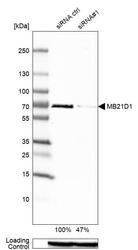
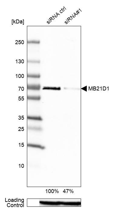
- Experimental details
- Western blot analysis in Rh30 cells transfected with control siRNA, target specific siRNA probe #1, using Anti-MB21D1 antibody. Remaining relative intensity is presented. Loading control: Anti-GAPDH.
Supportive validation
- Submitted by
- Atlas Antibodies (provider)
- Main image
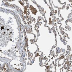
- Experimental details
- Immunohistochemical staining of human lung shows moderate to strong cytoplasmic positivity in macrophages and a subset of leukocytes.
- Sample type
- HUMAN

- Submitted by
- Atlas Antibodies (provider)
- Main image

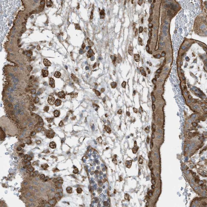
- Experimental details
- Immunohistochemical staining of human placenta shows moderate cytoplasmic positivity in Hofbauer cells and placental trophoblast.
- Sample type
- HUMAN
- Submitted by
- Atlas Antibodies (provider)
- Main image
- Experimental details
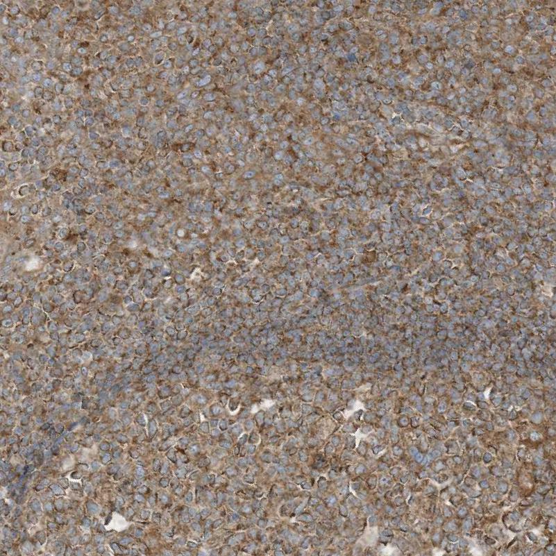
- Immunohistochemical staining of human tonsil shows moderate cytoplasmic positivity in lymphoid cells.
- Sample type
- HUMAN
- Submitted by
- Atlas Antibodies (provider)
- Main image
- Experimental details
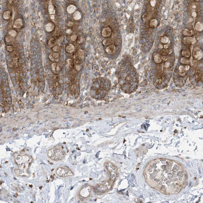
- Immunohistochemical staining of human duodenum shows moderate to strong cytoplasmic positivity in lymphoid cells in lamina propria.
- Sample type
- HUMAN
- Submitted by
- Atlas Antibodies (provider)
- Main image
- Experimental details
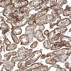
- Immunohistochemical staining of human placenta shows strong granular cytoplasmic positivity in trophoblastic cells.
- Sample type
- HUMAN

- Submitted by
- Atlas Antibodies (provider)
- Main image
- Experimental details
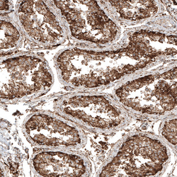
- Immunohistochemical staining of human testis shows strong granular cytoplasmic positivity in cells in seminiferous ducts.
- Sample type
- HUMAN

- Submitted by
- Atlas Antibodies (provider)
- Main image

- Experimental details
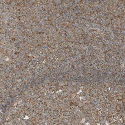
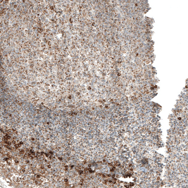
- Immunohistochemical staining of human tonsil shows moderate to strong granular cytoplasmic positivity in non-germinal center and germinal center cells.
- Sample type
- HUMAN
- Submitted by
- Atlas Antibodies (provider)
- Main image
- Experimental details
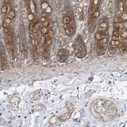
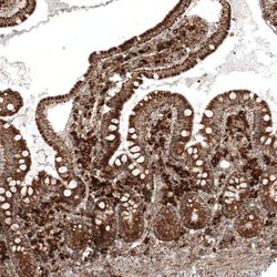
- Immunohistochemical staining of human small intestine shows strong granular cytoplasmic positivity in glandular cells.
- Sample type
- HUMAN
