Explore
 Validate
 Learn
Learn Immunocytochemistry
Antibody data
- Antibody Data
- Antigen structure
- References [0]
- Comments [0]
- Validations
- Immunocytochemistry [1]
- Immunohistochemistry [5]
Submit
Validation data
Reference
Comment
Report error
- Product number
- HPA031702 - Provider product page

- Provider
- Atlas Antibodies
- Proper citation
- Atlas Antibodies Cat#HPA031702, RRID:AB_2673999
- Product name
- Anti-MB21D1
- Antibody type
- Polyclonal
- Reactivity
- Human
- Host
- Rabbit
- Conjugate
- Unconjugated
- Antigen sequence
KHLDKFSSYHVKTAFFHVCTQNPQDSQWDRKDLGL
CFDNCVTYFLQCLRTEKLENYFIPEFNLFSSNLID
KRSKEFLTKQIEYER- Isotype
- IgG
- Vial size
- 100 µl
- Storage
- Store at +4°C for short term storage. Long time storage is recommended at -20°C.
No comments: Submit comment
Supportive validation
- Submitted by
- Atlas Antibodies (provider)
- Main image
- Experimental details
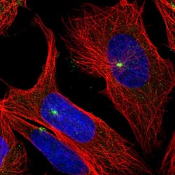
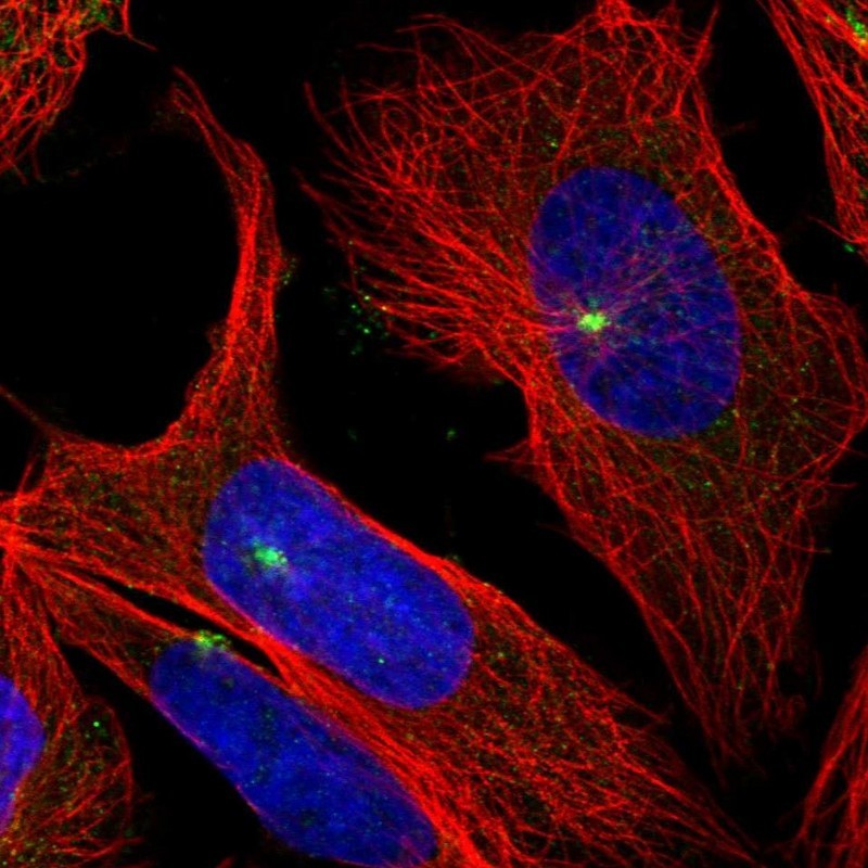
- Immunofluorescent staining of human cell line U-2 OS shows localization to microtubule organizing center.
- Sample type
- HUMAN
Supportive validation
- Submitted by
- Atlas Antibodies (provider)
- Main image
- Experimental details
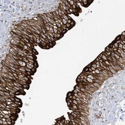
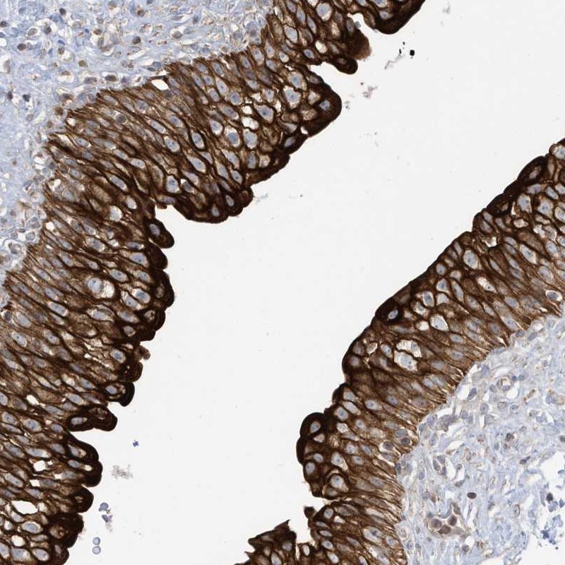
- Immunohistochemical staining of human urinary bladder shows strong cytoplasmic and membranous positivity in urothelial cells.
- Sample type
- HUMAN
- Submitted by
- Atlas Antibodies (provider)
- Main image
- Experimental details
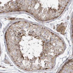
- Immunohistochemical staining of human testis shows moderate cytoplasmic positivity in cells in seminiferous ducts.
- Sample type
- HUMAN

- Submitted by
- Atlas Antibodies (provider)
- Main image
- Experimental details
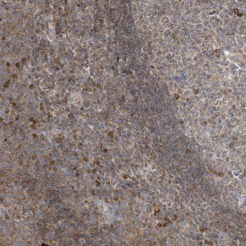
- Immunohistochemical staining of human tonsil shows moderate cytoplasmic positivity in non-germinal center cells.
- Sample type
- HUMAN
- Submitted by
- Atlas Antibodies (provider)
- Main image
- Experimental details
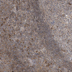
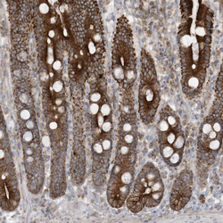
- Immunohistochemical staining of human upper gastrointestinal shows strong cytoplasmic positivity in glandular cells.
- Sample type
- HUMAN

- Submitted by
- Atlas Antibodies (provider)
- Main image
- Experimental details
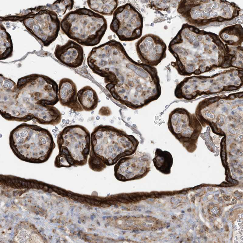
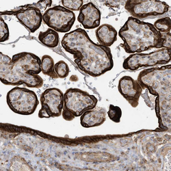
- Immunohistochemical staining of human placenta shows strong cytoplasmic positivity in trophoblastic cells.
- Sample type
- HUMAN