Explore
 Validate
 Learn
Learn Western blot
Antibody data
- Antibody Data
- Antigen structure
- References [0]
- Comments [0]
- Validations
- Western blot [2]
- Immunocytochemistry [2]
- Immunohistochemistry [12]
- Flow cytometry [1]
Submit
Validation data
Reference
Comment
Report error
- Product number
- TA500592 - Provider product page

- Provider
- OriGene
- Proper citation
- OriGene Cat#TA500592, RRID:AB_10585570
- Product name
- PPP5C (PP5) mouse monoclonal antibody, clone OTI4G8 (formerly 4G8)
- Antibody type
- Monoclonal
- Description
- PPP5C (PP5) mouse monoclonal antibody, clone OTI4G8 (formerly 4G8)
- Reactivity
- Canine
- Host
- Mouse
- Conjugate
- Unconjugated
- Epitope
- PPP5C
- Isotype
- IgG
- Antibody clone number
- OTI4G8
- Vial size
- 100 µl
- Concentration
- 0.9 mg/ml
No comments: Submit comment
Supportive validation
- Submitted by
- OriGene (provider)
- Main image
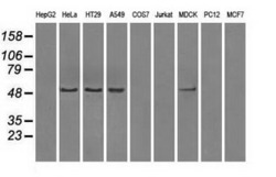
- Experimental details
- Western blot analysis of extracts (35ug) from 9 different cell lines by usin g anti-PPP5C monoclonal antibody (HepG2: human; HeLa: human; SVT2: mouse; A549: human; COS7: monkey; Jurkat: human; MDCK: canine; PC12: rat; MCF7: human).
- Validation comment
- WB

- Submitted by
- OriGene (provider)
- Main image
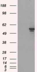
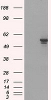
- Experimental details
- HEK293T cells were transfected with the pCMV6-ENTRY control (Left lane) or pCMV6-ENTRY PPP5C (RC201650, Right lane) cDNA for 48 hrs and lysed. Equivalent amounts of cell lysates (5 ug per lane) were separated by SDS-PAGE and immunoblotted with anti-PPP5C.
- Validation comment
- WB
Supportive validation
- Submitted by
- OriGene (provider)
- Main image
- Experimental details
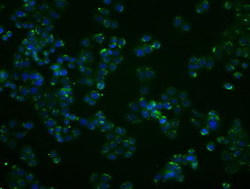
- Immunofluorescent staining of HT29 cells using anti-PPP5C mouse monoclonal antibody (TA500592).
- Validation comment
- IF

- Submitted by
- OriGene (provider)
- Main image

- Experimental details
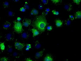
- Anti-PPP5C mouse monoclonal antibody (TA500592) immunofluorescent staining of COS7 cells transiently transfected by pCMV6-ENTRY PPP5C(RC201650).
- Validation comment
- IF
Supportive validation
- Submitted by
- OriGene (provider)
- Main image
- Experimental details
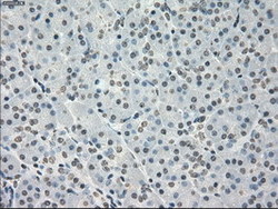
- Immunohistochemical staining of paraffin-embedded Human pancreas tissue within the normal limits using anti-PPP5C mouse monoclonal antibody. (Heat-induced epitope retrieval by 10mM citric buffer, pH6.0, 100C for 10min, TA500592)
- Validation comment
- IHC

- Submitted by
- OriGene (provider)
- Main image
- Experimental details
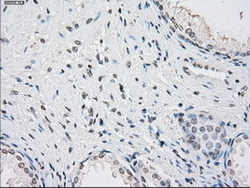
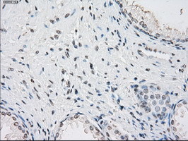
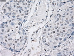
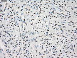
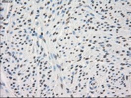
- Immunohistochemical staining of paraffin-embedded Human prostate tissue within the normal limits using anti-PPP5C mouse monoclonal antibody. (Heat-induced epitope retrieval by 10mM citric buffer, pH6.0, 100C for 10min, TA500592)
- Validation comment
- IHC
- Submitted by
- OriGene (provider)
- Main image
- Experimental details
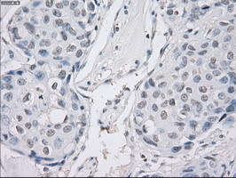
- Immunohistochemical staining of paraffin-embedded Human breast tissue within the normal limits using anti-PPP5C mouse monoclonal antibody. (Heat-induced epitope retrieval by 10mM citric buffer, pH6.0, 100C for 10min, TA500592)
- Validation comment
- IHC
- Submitted by
- OriGene (provider)
- Main image
- Experimental details
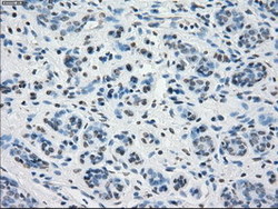
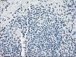
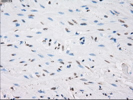
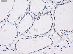
- Immunohistochemical staining of paraffin-embedded Carcinoma of Human thyroid tissue using anti-PPP5C mouse monoclonal antibody. (Heat-induced epitope retrieval by 10mM citric buffer, pH6.0, 100C for 10min, TA500592)
- Validation comment
- IHC
- Submitted by
- OriGene (provider)
- Main image
- Experimental details
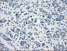
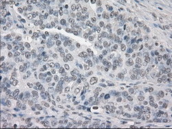
- Immunohistochemical staining of paraffin-embedded Adenocarcinoma of Human ovary tissue using anti-PPP5C mouse monoclonal antibody. (Heat-induced epitope retrieval by 10mM citric buffer, pH6.0, 100C for 10min, TA500592)
- Validation comment
- IHC

- Submitted by
- OriGene (provider)
- Main image
- Experimental details
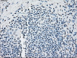
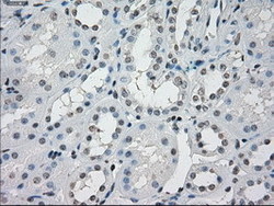
- Immunohistochemical staining of paraffin-embedded Human Kidney tissue within the normal limits using anti-PPP5C mouse monoclonal antibody. (Heat-induced epitope retrieval by 10mM citric buffer, pH6.0, 100C for 10min, TA500592)
- Validation comment
- IHC

- Submitted by
- OriGene (provider)
- Main image

- Experimental details
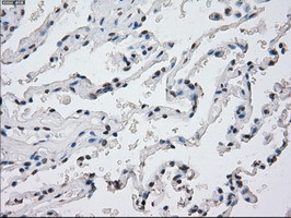
- Immunohistochemical staining of paraffin-embedded Human lung tissue within the normal limits using anti-PPP5C mouse monoclonal antibody. (Heat-induced epitope retrieval by 10mM citric buffer, pH6.0, 100C for 10min, TA500592)
- Validation comment
- IHC
- Submitted by
- OriGene (provider)
- Main image
- Experimental details
- Immunohistochemical staining of paraffin-embedded Adenocarcinoma of Human breast tissue using anti-PPP5C mouse monoclonal antibody. (Heat-induced epitope retrieval by 10mM citric buffer, pH6.0, 100C for 10min, TA500592)
- Validation comment
- IHC
- Submitted by
- OriGene (provider)
- Main image

- Experimental details
- Immunohistochemical staining of paraffin-embedded Adenocarcinoma of Human colon tissue using anti-PPP5C mouse monoclonal antibody. (Heat-induced epitope retrieval by 10mM citric buffer, pH6.0, 100C for 10min, TA500592)
- Validation comment
- IHC
- Submitted by
- OriGene (provider)
- Main image

- Experimental details
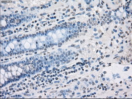
- Immunohistochemical staining of paraffin-embedded Human colon tissue within the normal limits using anti-PPP5C mouse monoclonal antibody. (Heat-induced epitope retrieval by 10mM citric buffer, pH6.0, 100C for 10min, TA500592)
- Validation comment
- IHC
- Submitted by
- OriGene (provider)
- Main image
- Experimental details
- Immunohistochemical staining of paraffin-embedded Human endometrium tissue within the normal limits using anti-PPP5C mouse monoclonal antibody. (Heat-induced epitope retrieval by 10mM citric buffer, pH6.0, 100C for 10min, TA500592)
- Validation comment
- IHC
- Submitted by
- OriGene (provider)
- Main image
- Experimental details
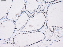
- Immunohistochemical staining of paraffin-embedded Human thyroid tissue within the normal limits using anti-PPP5C mouse monoclonal antibody. (Heat-induced epitope retrieval by 10mM citric buffer, pH6.0, 100C for 10min, TA500592)
- Validation comment
- IHC
Supportive validation
- Submitted by
- OriGene (provider)
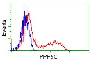
- Main image
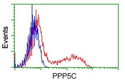
- Experimental details
- HEK293T cells transfected with either RC201650 overexpress plasmid(Red) or empty vector control plasmid(Blue) were immunostained by anti-PPP5C antibody(TA500592), and then analyzed by flow cytometry.
- Validation comment
- FC