Explore
 Validate
 Learn
Learn Western blot
 Immunohistochemistry
Antibody data
- Antibody Data
- Antigen structure
- References [1]
- Comments [0]
- Validations
- Immunohistochemistry [1]
Submit
Validation data
Reference
Comment
Report error
- Product number
- AF3067 - Provider product page

- Provider
- R&D Systems
- Product name
- Mouse SorCS3 Antibody
- Antibody type
- Polyclonal
- Description
- Immunogen affinity purified. Detects mouse SorCS3 in direct ELISAs and Western blots. In direct ELISAs, approximately 40% cross-reactivity with recombinant human SorCS3 is observed, approximately 7% cross-reactivity with recombinant mouse (rm) SorCS1 is observed, and less than 2% cross-reactivity with rmSorCS2 is observed.
- Reactivity
- Mouse
- Host
- Goat
- Conjugate
- Unconjugated
- Antigen sequence
Q8VI51- Isotype
- IgG
- Vial size
- 100 ug
- Concentration
- LYOPH
- Storage
- Use a manual defrost freezer and avoid repeated freeze-thaw cycles. 12 months from date of receipt, -20 to -70 °C as supplied. 1 month, 2 to 8 °C under sterile conditions after reconstitution. 6 months, -20 to -70 °C under sterile conditions after reconstitution.
Submitted references Tbr1 instructs laminar patterning of retinal ganglion cell dendrites.
Liu J, Reggiani JDS, Laboulaye MA, Pandey S, Chen B, Rubenstein JLR, Krishnaswamy A, Sanes JR
Nature neuroscience 2018 May;21(5):659-670
Nature neuroscience 2018 May;21(5):659-670
No comments: Submit comment
Supportive validation
- Submitted by
- R&D Systems (provider)
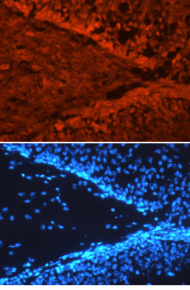
- Main image

- Experimental details
- SorCS3 in Mouse Brain. SorCS3 was detected in immersion fixed frozen sections of mouse brain (hippocampus) using 10 µg/mL Goat Anti-Mouse SorCS3 Antigen Affinity-purified Polyclonal Antibody (Catalog # AF3067) overnight at 4 °C. Tissue was stained with the NorthernLights™ 557-conjugated Anti-Goat IgG Secondary Antibody (red, upper panel; Catalog # NL001) and counterstained with DAPI (blue, lower panel). View our protocol for Fluorescent IHC Staining of Frozen Tissue Sections.