 Explore
 Validate
 Learn
Learn Western blot
 Immunoprecipitation
Antibody data
- Antibody Data
- Antigen structure
- References [5]
- Comments [0]
- Validations
- Western blot [2]
- Immunohistochemistry [1]
Submit
Validation data
Reference
Comment
Report error
- Product number
- NB300-206 - Provider product page

- Provider
- Novus Biologicals
- Proper citation
- Novus Cat#NB300-206, RRID:AB_10002355
- Product name
- Mouse Monoclonal HIP1 Related Antibody
- Antibody type
- Monoclonal
- Description
- Unpurified.
- Reactivity
- Human, Mouse, Rat, Bacteria
- Host
- Mouse
- Isotype
- IgG
- Vial size
- 0.1 ml
- Storage
- Aliquot and store at -20C or -80C. Avoid freeze-thaw cycles.
Submitted references DEAD-box helicase DDX27 regulates 3' end formation of ribosomal 47S RNA and stably associates with the PeBoW-complex.
Huntingtin interacting protein 1 modulates the transcriptional activity of nuclear hormone receptors.
Hip1-related mutant mice grow and develop normally but have accelerated spinal abnormalities and dwarfism in the absence of HIP1.
Hip1-related mutant mice grow and develop normally but have accelerated spinal abnormalities and dwarfism in the absence of HIP1.
HIP1 and HIP1r stabilize receptor tyrosine kinases and bind 3-phosphoinositides via epsin N-terminal homology domains.
Kellner M, Rohrmoser M, Forné I, Voss K, Burger K, Mühl B, Gruber-Eber A, Kremmer E, Imhof A, Eick D
Experimental cell research 2015 May 15;334(1):146-59
Experimental cell research 2015 May 15;334(1):146-59
Huntingtin interacting protein 1 modulates the transcriptional activity of nuclear hormone receptors.
Mills IG, Gaughan L, Robson C, Ross T, McCracken S, Kelly J, Neal DE
The Journal of cell biology 2005 Jul 18;170(2):191-200
The Journal of cell biology 2005 Jul 18;170(2):191-200
Hip1-related mutant mice grow and develop normally but have accelerated spinal abnormalities and dwarfism in the absence of HIP1.
Hyun TS, Li L, Oravecz-Wilson KI, Bradley SV, Provot MM, Munaco AJ, Mizukami IF, Sun H, Ross TS
Molecular and cellular biology 2004 May;24(10):4329-40
Molecular and cellular biology 2004 May;24(10):4329-40
HIP1 and HIP1r stabilize receptor tyrosine kinases and bind 3-phosphoinositides via epsin N-terminal homology domains.
Hyun TS, Rao DS, Saint-Dic D, Michael LE, Kumar PD, Bradley SV, Mizukami IF, Oravecz-Wilson KI, Ross TS
The Journal of biological chemistry 2004 Apr 2;279(14):14294-306
The Journal of biological chemistry 2004 Apr 2;279(14):14294-306
No comments: Submit comment
Supportive validation
Supportive validation
- Submitted by
- Wang Huanhuan
- Main image

- Experimental details
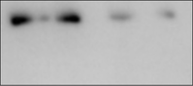
- Whole cell lysis from Neuro2a cells and 293ft transfected with Hip1r-HA-tag(harvest at 48h after transfection), WB follow normal protocols
- Sample type
- Neuro2a is a neuroblastoma cell line from mouse, 293ft is a human cell line
- Validation comment
- Hip1r protein is above 100KDa, the band is specific at above 100Kda, and get same band at HA-tagged protein wihch is validated by sequence after clone
- Primary Ab dilution
- 1:1000
- Secondary Ab
- Secondary Ab
- Secondary Ab dilution
- 1:5000
Supportive validation
- Submitted by
- Novus Biologicals (provider)
- Main image
- Experimental details
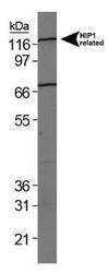
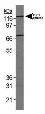
- Western Blot: HIP1 Related Antibody (1E1) [NB300-206] - Analysis of HIP1 Related on HeLa whole cell extract.
Supportive validation
- Submitted by
- Novus Biologicals (provider)
- Main image

- Experimental details
- Immunohistochemistry-Paraffin: HIP1 Related Antibody (1E1) [NB300-206] - Analysis of a FFPE tissue section of human kidney using 1:200 concentration of HIP1 Related antibody. The staining was developed using HRP labeled anti-rabbit secondary antibody and DAB reagent, and nuclei of cells were counter-stained with hematoxylin. This HIP1 related antibody generated strong staining of the cytoplasm and membrane.
