 Explore
 Validate
 Learn
Learn Immunocytochemistry
Antibody data
- Antibody Data
- Antigen structure
- References [2]
- Comments [0]
- Validations
- Immunocytochemistry [1]
- Immunohistochemistry [5]
Submit
Validation data
Reference
Comment
Report error
- Product number
- HPA041314 - Provider product page

- Provider
- Atlas Antibodies
- Proper citation
- Atlas Antibodies Cat#HPA041314, RRID:AB_10795587
- Product name
- Anti-RILPL1
- Antibody type
- Polyclonal
- Reactivity
- Human
- Host
- Rabbit
- Conjugate
- Unconjugated
- Antigen sequence
LELDRLRLERMDRIEKERKHQKELELVEDVWRGEA
QDLLSQIAQLQEENKQLMTNLSHKDVNFSEEEFQK
HEGMSERERQVMKKLKEV- Isotype
- IgG
- Vial size
- 100 µl
- Storage
- Store at +4°C for short term storage. Long time storage is recommended at -20°C.
Submitted references The Rilp-like proteins Rilpl1 and Rilpl2 regulate ciliary membrane content.
The interactome of LIM domain proteins: The contributions of LIM domain proteins to heart failure and heart development
Schaub JR, Stearns T
Molecular biology of the cell 2013 Feb;24(4):453-64
Molecular biology of the cell 2013 Feb;24(4):453-64
The interactome of LIM domain proteins: The contributions of LIM domain proteins to heart failure and heart development
Li A, Ponten F, dos Remedios C
PROTEOMICS 2012 January;12(2):203-225
PROTEOMICS 2012 January;12(2):203-225
No comments: Submit comment
Supportive validation
- Submitted by
- Atlas Antibodies (provider)
- Main image

- Experimental details
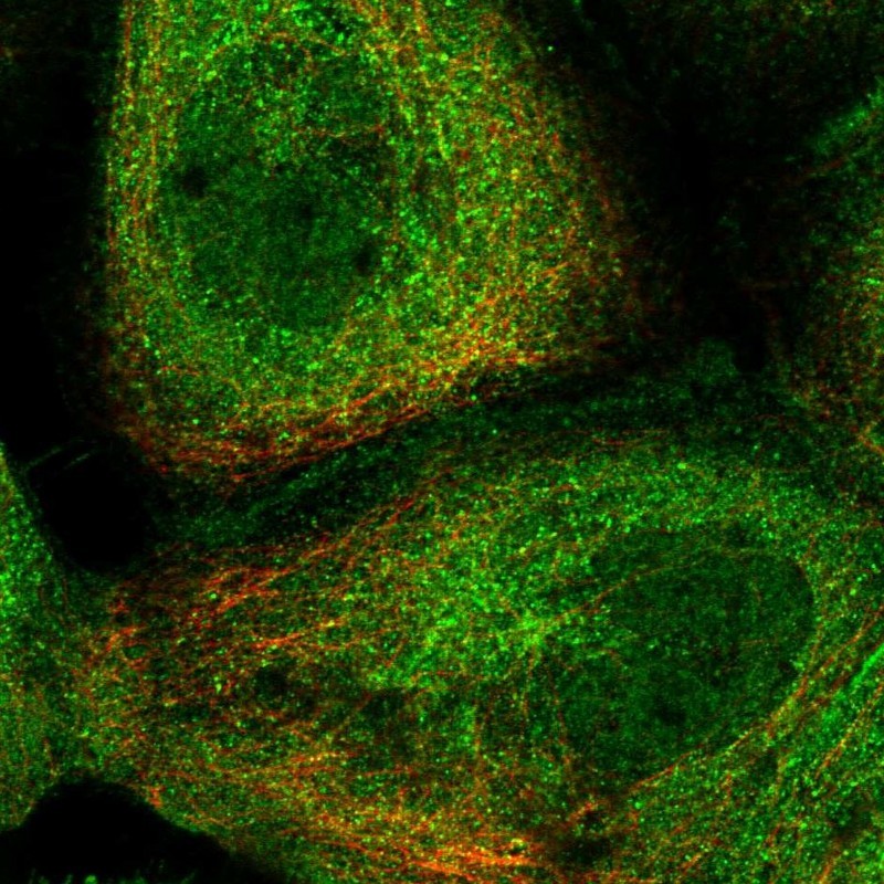
- Immunofluorescent staining of human cell line A-431 shows localization to nucleoplasm, plasma membrane & cytosol.
- Sample type
- HUMAN
Supportive validation
- Submitted by
- Atlas Antibodies (provider)
- Main image

- Experimental details
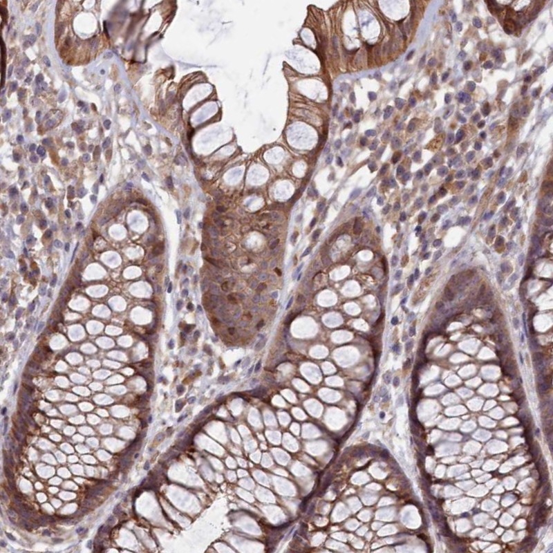
- Immunohistochemical staining of human rectum shows moderate cytoplasmic positivity in glandular cells.
- Submitted by
- Atlas Antibodies (provider)
- Main image

- Experimental details
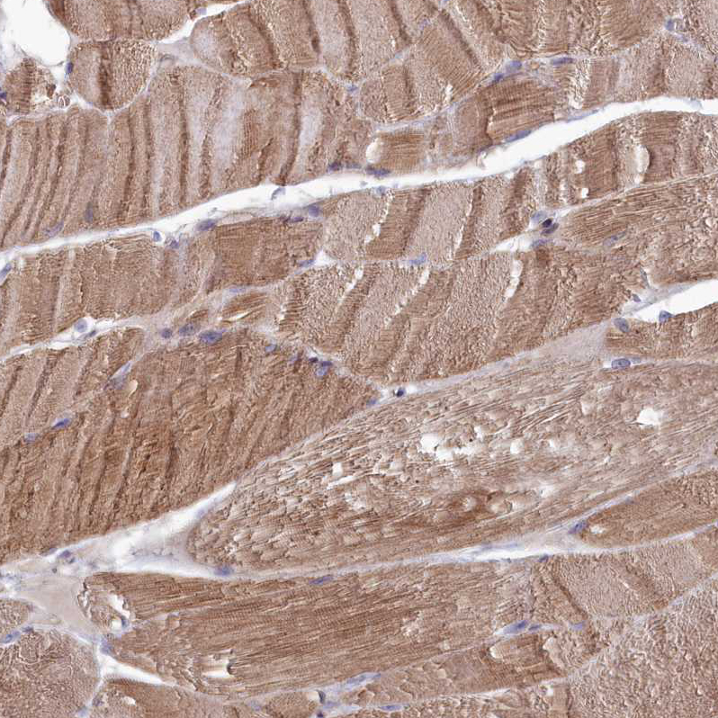
- Immunohistochemical staining of human skeletal muscle shows moderate cytoplasmic positivity in myocytes.
- Sample type
- HUMAN
- Submitted by
- Atlas Antibodies (provider)
- Main image

- Experimental details
- Immunohistochemical staining of human skin shows weak cytoplasmic positivity in squamous epithelial cells.
- Sample type
- HUMAN

- Submitted by
- Atlas Antibodies (provider)
- Main image

- Experimental details
- Immunohistochemical staining of human prostate shows moderate cytoplasmic positivity in smooth muscle cells and weak membranous staining of glandular cells.
- Sample type
- HUMAN

- Submitted by
- Atlas Antibodies (provider)
- Main image

- Experimental details
- Immunohistochemical staining of human placenta shows strong cytoplasmic positivity in trophoblastic cells.
- Sample type
- HUMAN
