Explore
 Validate
 Learn
LearnBS60737
antibody from Bioworld Technology, Inc
Targeting: ATP6V0A2
a2, ATP6a2, ATP6N1D, J6B7, Stv1, TJ6, TJ6M, TJ6s, Vph1
 Western blot
Antibody data
- Antibody Data
- Antigen structure
- References [0]
- Comments [0]
- Validations
- Western blot [1]
Submit
Validation data
Reference
Comment
Report error
- Product number
- BS60737 - Provider product page

- Provider
- Bioworld Technology, Inc
- Product name
- ATP6V0A2 polyclonal antibody
- Antibody type
- Polyclonal
- Antigen
- A synthetic peptide corresponding to residues in Human ATP6V0A2.
- Description
- Vacuolar-type H+-ATPase (V-ATPase) is a multisubunit enzyme responsible for the acidification of eukaryotic intracellular organelles. V-ATPases pump protons against an electrochemical gradient, while F-ATPases reverse the process, thereby synthesizing ATP. A peripheral V1 domain, which is responsible for ATP hydrolysis, and an integral V0 domain, which is responsible for proton translocation, comprise the V-ATPase complex. Nine subunits (AÿH) make up the V1 domain and five subunits (A, D, C, C' and C") make up the V0 domain. As part of the V0 domain, V-ATPase A2 (ATPase, H+ transporting, lysosomal V0 subunit a2), consists of 856 amino acids and is also known as ATP6V0A2, V-type proton ATPase subunit a isoform 2, vacuolar proton translocating ATPase subunit a isoform 2, lysosomal H(+)-transporting ATPase V0 subunit a2 or TJ6. V-ATPase A2 is a multi-pass membrane protein with localization in the cell membrane, endosome membrane and the subapical vesicles of the kidney£ªs proximal tubules. V-ATPase A2 plays an important role in Golgi function by regulating pH. Wrinkly skin syndrome (WSS) and cutis laxa type II (ARCL type II) are caused as a result of V-ATPase A2 defects.
- Reactivity
- Human, Mouse
- Host
- Rabbit
- Isotype
- IgG
- Vial size
- 100ul
- Concentration
- 1 mg/ml
- Storage
- Store at 4°C short term. Aliquot and store at -27°C long term. Avoid freeze-thaw cycles.
No comments: Submit comment
Supportive validation
- Submitted by
- Bioworld Technology, Inc (provider)
- Main image
- Experimental details
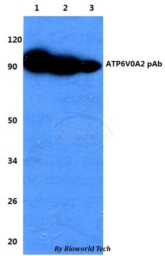
- Western blot (WB) analysis of ATP6V0A2 pAb at 1:500 dilutionLane1:HEK293T whole cell lysateLane2:sp2/0 whole cell lysateLane3:H9C9 whole cell lysate