Explore
 Validate
 Learn
Learn Western blot
Antibody data
- Antibody Data
- Antigen structure
- References [1]
- Comments [0]
- Validations
- Western blot [1]
- Immunohistochemistry [1]
Submit
Validation data
Reference
Comment
Report error
- Product number
- MAB7509 - Provider product page

- Provider
- R&D Systems
- Product name
- Human CHMP2B Antibody
- Antibody type
- Monoclonal
- Description
- Protein A or G purified from hybridoma culture supernatant. Detects human CHMP2B in direct ELISAs and Western blots
- Reactivity
- Human
- Host
- Mouse
- Conjugate
- Unconjugated
- Antigen sequence
Q9UQN3- Isotype
- IgG
- Antibody clone number
- 791521
- Vial size
- 100 ug
- Concentration
- LYOPH
- Storage
- Use a manual defrost freezer and avoid repeated freeze-thaw cycles. 12 months from date of receipt, -20 to -70 °C as supplied. 1 month, 2 to 8 °C under sterile conditions after reconstitution. 6 months, -20 to -70 °C under sterile conditions after reconstitution.
Submitted references The ESCRT-Related ATPase Vps4 Is Modulated by Interferon during Herpes Simplex Virus 1 Infection.
Cabrera JR, Manivanh R, North BJ, Leib DA
mBio 2019 Mar 5;10(2)
mBio 2019 Mar 5;10(2)
No comments: Submit comment
Supportive validation
- Submitted by
- R&D Systems (provider)
- Main image
- Experimental details
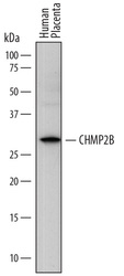
- Detection of Human CHMP2B by Western Blot. Western blot shows lysates of human placenta tissue. PVDF membrane was probed with 2 µg/mL of Mouse Anti-Human CHMP2B Monoclonal Antibody (Catalog # MAB7509) followed by HRP-conjugated Anti-Mouse IgG Secondary Antibody (Catalog # HAF018). A specific band was detected for CHMP2B at approximately 30 kDa (as indicated). This experiment was conducted under reducing conditions and using Immunoblot Buffer Group 1.

Supportive validation
- Submitted by
- R&D Systems (provider)
- Main image

- Experimental details
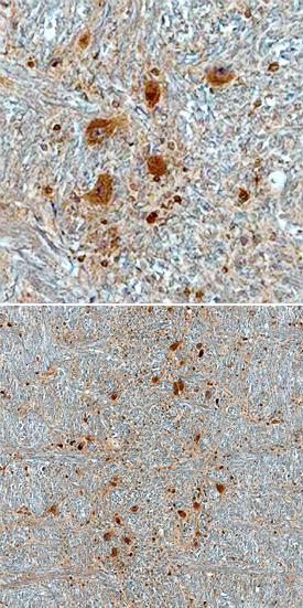
- CHMP2B in Human Brain. CHMP2B was detected in immersion fixed paraffin-embedded sections of human brain (medulla) using Mouse Anti-Human CHMP2B Monoclonal Antibody (Catalog # MAB7509) at 3 µg/mL overnight at 4 °C. Before incubation with the primary antibody, tissue was subjected to heat-induced epitope retrieval using Antigen Retrieval Reagent-Basic (Catalog # CTS013). Tissue was stained using the Anti-Mouse HRP-DAB Cell & Tissue Staining Kit (brown; Catalog # CTS002) and counterstained with hematoxylin (blue). Staining is shown at both high (upper panel) and low (lower panel) magnification, and specific staining was localized to neuronal cytoplasm. View our protocol for Chromogenic IHC Staining of Paraffin-embedded Tissue Sections.