 Explore
 Validate
 Learn
Learn Western blot
 ELISA
ELISAAntibody data
- Antibody Data
- Antigen structure
- References [0]
- Comments [0]
- Validations
- Western blot [3]
- Immunohistochemistry [1]
- Flow cytometry [1]
Submit
Validation data
Reference
Comment
Report error
- Product number
- NB110-55454 - Provider product page

- Provider
- Novus Biologicals
- Proper citation
- Novus Cat#NB110-55454, RRID:AB_837588
- Product name
- Mouse Monoclonal Apolipoprotein A5 Antibody
- Antibody type
- Monoclonal
- Description
- Protein G purified.
- Reactivity
- Human
- Host
- Mouse
- Isotype
- IgG
- Vial size
- 0.1 ml
- Concentration
- 1.9 mg/ml
- Storage
- Aliquot and store at -20C or -80C. Avoid freeze-thaw cycles.
No comments: Submit comment
Supportive validation
- Submitted by
- Novus Biologicals (provider)
- Main image

- Experimental details
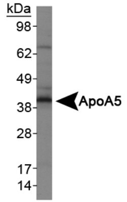
- Western Blot: Apolipoprotein A5 Antibody (1G5G9) [NB110-55454] - Analysis of ApoA5 in HepG2 lysates
- Submitted by
- Novus Biologicals (provider)
- Main image
- Experimental details
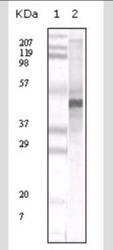
- Western Blot: Apolipoprotein A5 Antibody (1G5G9) [NB110-55454] - Analysis in human serum total protein (lane 2). Lane 1 is a MW standard.

- Submitted by
- Novus Biologicals (provider)
- Main image

- Experimental details
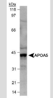
- Western Blot: Apolipoprotein A5 Antibody (1G5G9) [NB110-55454] - Detection of Apo A5 in HepG2 cell lysate.
Supportive validation
- Submitted by
- Novus Biologicals (provider)
- Main image
- Experimental details
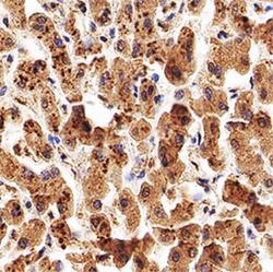
- Immunohistochemistry-Paraffin: Apolipoprotein A5 Antibody (1G5G9) [NB110-55454] - IHC analysis of a formalin fixed paraffin-embedded (FFPE) human liver using 5 ug/ml conc. of Apolipoprotein A5 antibody on a Bond Rx autostainer (Leica Biosystems). The assay involved 20 minutes of heat induced antigen retrieval (HIER) using 10mM sodium citrate buffer (pH 6.0) and endogenous peroxidase quenching with peroxide block. The sections were incubated with primary antibody for 30 minutes and Bond Polymer Refine Detection (Leica Biosystems) with DAB was used for signal development followed by counterstaining with hematoxylin. Whole slide scanning and capturing of representative images was performed using Aperio AT2 (Leica Biosystems). Cytoplasmic staining of Apolipoprotein A5 in hepatocytes was observed. Staining was performed by Histowiz.

Supportive validation
- Submitted by
- Novus Biologicals (provider)
- Main image

- Experimental details
- Flow Cytometry: Apolipoprotein A5 Antibody (1G5G9) [NB110-55454] - Intracellular flow cytometric staining of 1 x 10^6 CHO (A) and HEK-293 (B) cells using Apolipoprotein A5 antibody (dark blue). Isotype control shown in orange. An antibody concentration of 1 ug/1x10^6 cells was used.
