Explore
 Validate
 Learn
Learn Western blot
Antibody data
- Antibody Data
- Antigen structure
- References [1]
- Comments [0]
- Validations
- Western blot [5]
- Immunocytochemistry [1]
- Immunohistochemistry [1]
- Flow cytometry [1]
Submit
Validation data
Reference
Comment
Report error
- Product number
- MA1-46001 - Provider product page

- Provider
- Invitrogen Antibodies
- Product name
- CIP2A Monoclonal Antibody (2G10)
- Antibody type
- Monoclonal
- Antigen
- Other
- Description
- Suggested positive control: Hela whole cell extract, antigen standard for KIAA1524 (transient overexpression lysate).
- Reactivity
- Human, Mouse, Rat
- Host
- Mouse
- Isotype
- IgG
- Antibody clone number
- 2G10
- Vial size
- 100 µL
- Concentration
- 1.0 mg/mL
- Storage
- -20° C, Avoid Freeze/Thaw Cycles
Submitted references Diane-35 and Metformin Induce Autophagy and Apoptosis in Polycystic Ovary Syndrome Women with Early-Stage Endometrial Carcinoma.
Liu Y, Wang Y, Yao D, Chen X, Zhang F, Feng Y, Li X
Genes 2022 Jan 12;13(1)
Genes 2022 Jan 12;13(1)
No comments: Submit comment
Supportive validation
- Submitted by
- Invitrogen Antibodies (provider)
- Main image

- Experimental details
- Western Blot detection of p90 autoantigen in HeLa whole cell lysate
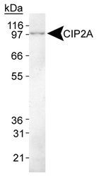
- Submitted by
- Invitrogen Antibodies (provider)
- Main image

- Experimental details
- Western blot analysis of CIP2A using a monoclonal antibody (Product # MA1-46001).

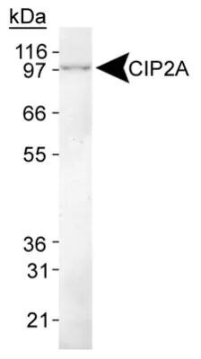
- Submitted by
- Invitrogen Antibodies (provider)
- Main image

- Experimental details
- Western blot analysis of CIP2A in HeLa whole cell lysate. Sample was incubated in CIP2A monoclonal antibody (Product # MA1-46001).
- Submitted by
- Invitrogen Antibodies (provider)
- Main image

- Experimental details
- Western blot analysis of CIP2A in NIH/3T3 cell lysate. Sample was incubated in CIP2A monoclonal antibody (Product # MA1-46001).

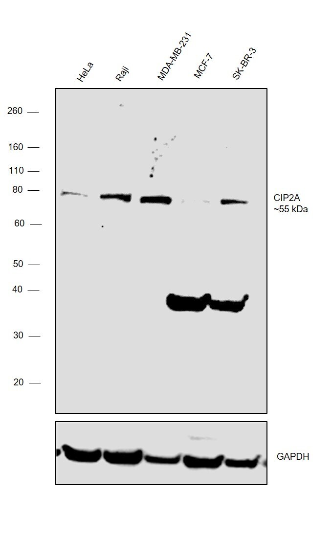
- Submitted by
- Invitrogen Antibodies (provider)
- Main image

- Experimental details
- Western blot was performed using Anti- CIP2A Monoclonal Antibody (Product # MA1-46001) and an ~55kDa band corresponding to CIP2A was observed across the cell lines tested except for MCF-7 which is reported to be a low expressor for CIP2A. An uncharacterized band was also observed in MCF-7 and SK-BR-3 cells at ~35kDa. Whole cell extracts (30 µg lysate) of HeLa (Lane 1), Raji (Lane 2), MDA-MB-231 (Lane 3), MCF-7 (Lane 4) and SK-BR-3 (Lane 5) were electrophoresed using NuPAGE™ 4-12% Bis-Tris Protein Gel (Product # NP0321BOX). Resolved proteins were then transferred onto a nitrocellulose membrane (Product # IB23001) by iBlot® 2 Dry Blotting System (Product # IB21001). The blot was probed with the primary antibody (1:1000 dilution ) and detected by chemiluminescence with Goat anti-Mouse IgG (H+L), Superclonal™ Recombinant Secondary Antibody, HRP (Product # A28177, 1:4000 dilution) using the iBright FL 1000 (Product # A32752). Chemiluminescent detection was performed using Novex® ECL Chemiluminescent Substrate Reagent Kit (Product # WP20005).
Supportive validation
- Submitted by
- Invitrogen Antibodies (provider)
- Main image
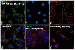
- Experimental details
- Immunofluorescence analysis of CIP2A was performed using 70% confluent log phase MDA-MB-231 cells. The cells were fixed with 4% paraformaldehyde for 10 minutes, permeabilized with 0.1% Triton™ X-100 for 15 minutes, and blocked with 2% BSA for 1 hour at room temperature. The cells were labeled with CIP2A Monoclonal Antibody (2G10) (Product # MA1-46001) at 1:200 dilution in 0.1% BSA, incubated at 4 degree celsius overnight and then with Goat anti- Mouse IgG (H+L) Highly Cross-Adsorbed Secondary Antibody, Alexa Fluor Plus 488 (Product # A28175) at a dilution of 1:2000 for 45 minutes at room temperature (Panel a: green). Nuclei (Panel b: blue) were stained with ProLong™ Diamond Antifade Mountant with DAPI (Product # P36962). F-actin (Panel c: red) was stained with Rhodamine Phalloidin (Product # R415, 1:300). Panel d represents the merged image showing localization in cytoplasm. Panel e represents low expressing cell line MCF7 with no expression. Panel f represents control cells with no primary antibody to assess background. The images were captured at 60X magnification

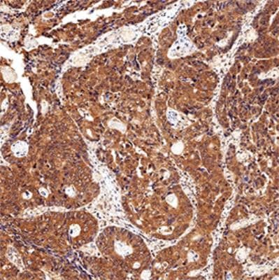
Supportive validation
- Submitted by
- Invitrogen Antibodies (provider)
- Main image

- Experimental details
- Immunohistochemical analysis of CIP2A in formalin fixed paraffin-embedded (FFPE) human colon cancer. Samples were incubated in CIP2A monoclonal antibody (Product # MA1-46001) using a dilution of 1:200. Bond Rx autostainer (Leica Biosystems). The assay involved 20 minutes of heat induced antigen retrieval (HIER) using 10mM sodium citrate buffer (pH 6.0) and endogenous peroxidase quenching with peroxide block. The sections were incubated with primary antibody for 30 minutes and Bond Polymer Refine Detection (Leica Biosystems) with DAB was used for signal development followed by counterstaining with hematoxylin. Whole slide scanning and capturing of representative images was performed using Aperio AT2 (Leica Biosystems). Staining was performed by Histowiz.
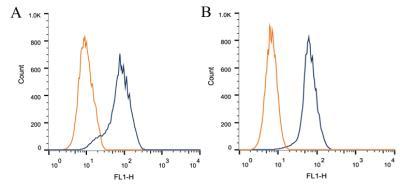
Supportive validation
- Submitted by
- Invitrogen Antibodies (provider)
- Main image
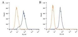
- Experimental details
- Flow cytometry of CIP2A in 1 x 10^6 CHO (A) and HEK-293 (B) cells. Samples were incubated in CIP2A monoclonal antibody (Product # MA1-46001) using a dilution of 1 µg/1x10^6 cells. Antibody (dark blue). Isotype control shown in orange.