 Explore
 Validate
 Learn
Learn Western blot
Antibody data
- Antibody Data
- Antigen structure
- References [5]
- Comments [0]
- Validations
- Western blot [4]
- Immunocytochemistry [3]
- Immunohistochemistry [2]
Submit
Validation data
Reference
Comment
Report error
- Product number
- GTX102255 - Provider product page

- Provider
- GeneTex
- Proper citation
- GeneTex Cat#GTX102255, RRID:AB_1950534
- Product name
- ORP150 antibody [C2C3], C-term
- Antibody type
- Polyclonal
- Reactivity
- Human, Mouse, Rat
- Host
- Rabbit
Submitted references PERK Signaling Regulates Extracellular Proteostasis of an Amyloidogenic Protein During Endoplasmic Reticulum Stress.
Elevated chaperone proteins are a feature of winter freeze avoidance by larvae of the goldenrod gall moth, Epiblema scudderiana.
The nucleotide exchange factors Grp170 and Sil1 induce cholera toxin release from BiP to enable retrotranslocation.
Unfolded protein response-induced ERdj3 secretion links ER stress to extracellular proteostasis.
Stress-independent activation of XBP1s and/or ATF6 reveals three functionally diverse ER proteostasis environments.
Romine IC, Wiseman RL
Scientific reports 2019 Jan 23;9(1):410
Scientific reports 2019 Jan 23;9(1):410
Elevated chaperone proteins are a feature of winter freeze avoidance by larvae of the goldenrod gall moth, Epiblema scudderiana.
Zhang G, Storey JM, Storey KB
Journal of insect physiology 2018 Apr;106(Pt 2):106-113
Journal of insect physiology 2018 Apr;106(Pt 2):106-113
The nucleotide exchange factors Grp170 and Sil1 induce cholera toxin release from BiP to enable retrotranslocation.
Williams JM, Inoue T, Chen G, Tsai B
Molecular biology of the cell 2015 Jun 15;26(12):2181-9
Molecular biology of the cell 2015 Jun 15;26(12):2181-9
Unfolded protein response-induced ERdj3 secretion links ER stress to extracellular proteostasis.
Genereux JC, Qu S, Zhou M, Ryno LM, Wang S, Shoulders MD, Kaufman RJ, Lasmézas CI, Kelly JW, Wiseman RL
The EMBO journal 2015 Jan 2;34(1):4-19
The EMBO journal 2015 Jan 2;34(1):4-19
Stress-independent activation of XBP1s and/or ATF6 reveals three functionally diverse ER proteostasis environments.
Shoulders MD, Ryno LM, Genereux JC, Moresco JJ, Tu PG, Wu C, Yates JR 3rd, Su AI, Kelly JW, Wiseman RL
Cell reports 2013 Apr 25;3(4):1279-92
Cell reports 2013 Apr 25;3(4):1279-92
No comments: Submit comment
Supportive validation
- Submitted by
- GeneTex (provider)
- Main image
- Experimental details
- Sample(30 ?g of whole cell lysate)A:H12997.5% SDS PAGEGTX102255 diluted at 1:1000
- Validation comment
- WB

- Submitted by
- GeneTex (provider)
- Main image

- Experimental details
- ORP150 antibody detects ORP150 protein by western blot analysis. Mouse tissue extracts (50 ?g) was separated by 5% SDS-PAGE, and blotted with ORP150 antibody (GTX102255) diluted by 1:500. The HRP-conjugated anti-rabbit IgG antibody (GTX213110-01) was used to detect the primary antibody.

- Submitted by
- GeneTex (provider)
- Main image

- Experimental details
- ORP150 antibody detects ORP150 protein by western blot analysis. Rat tissue extracts (50 ?g) was separated by 5% SDS-PAGE, and blotted with ORP150 antibody (GTX102255) diluted by 1:1000. The HRP-conjugated anti-rabbit IgG antibody (GTX213110-01) was used to detect the primary antibody.

- Submitted by
- GeneTex (provider)
- Main image

- Experimental details
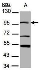
- ORP150 antibody detects ORP150 protein by western blot analysis. Various whole cell extracts (30 ?g) were separated by 5% SDS-PAGE, and blotted with ORP150 antibody (GTX102255) diluted by 1:1000. The HRP-conjugated anti-rabbit IgG antibody (GTX213110-01) was used to detect the primary antibody.

Supportive validation
- Submitted by
- GeneTex (provider)
- Main image

- Experimental details
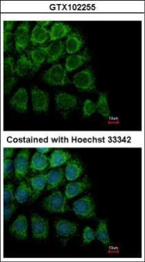
- Immunofluorescence analysis of methanol-fixed Hep3B, using ORP150(GTX102255) antibody at 1:500 dilution.
- Submitted by
- GeneTex (provider)
- Main image

- Experimental details
- ORP150 antibody [C2C3], C-term detects ORP150 protein at endoplasmic reticulum by immunofluorescent analysis.Sample: HeLa cells were fixed in 4% paraformaldehyde at RT for 15 min.Green: ORP150 protein stained by ORP150 antibody [C2C3], C-term (GTX102255) diluted at 1:200.Red: phalloidin, a cytoskeleton marker, stained by phalloidin (invitrogen, A12380) diluted at 1:200.Blue: Hoechst 33342 staining.

- Submitted by
- GeneTex (provider)
- Main image

- Experimental details
- ORP150 antibody [C2C3], C-term detects ORP150 protein at cytoplasm by immunofluorescent analysis.Sample: U2OS cells were fixed in 4% paraformaldehyde at RT for 15 min.Green: ORP150 protein stained by ORP150 antibody [C2C3], C-term (GTX102255) diluted at 1:1000.Red: alpha Tubulin, a cytoskeleton marker, stained by alpha Tubulin antibody [GT114] (GTX628802) diluted at 1:1000.Blue: Hoechst 33342 staining.

Supportive validation
- Submitted by
- GeneTex (provider)
- Main image
- Experimental details
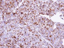
- Immunohistochemical analysis of paraffin-embedded CL1-0 xenograft, using ORP150(GTX102255) antibody at 1:100 dilution.

- Submitted by
- GeneTex (provider)
- Main image

- Experimental details
- ORP150 antibody [C2C3], C-term detects ORP150 protein at cytoplasm on mouse fore brain by immunohistochemical analysis. Sample: Paraffin-embedded mouse fore brain. ORP150 antibody [C2C3], C-term (GTX102255) diluted at 1:500.
