 Explore
 Validate
 Learn
Learn Western blot
Antibody data
- Antibody Data
- Antigen structure
- References [1]
- Comments [0]
- Validations
- Western blot [5]
- Immunocytochemistry [3]
- Immunohistochemistry [2]
Submit
Validation data
Reference
Comment
Report error
- Product number
- PA5-27655 - Provider product page

- Provider
- Invitrogen Antibodies
- Product name
- HYOU1 Polyclonal Antibody
- Antibody type
- Polyclonal
- Antigen
- Recombinant protein fragment
- Description
- Recommended positive controls: MCF-7, MDA-MB-231, mouse liver, rat brain.
- Concentration
- 0.41 mg/mL
Submitted references ERp18 regulates activation of ATF6α during unfolded protein response.
Oka OB, van Lith M, Rudolf J, Tungkum W, Pringle MA, Bulleid NJ
The EMBO journal 2019 Aug 1;38(15):e100990
The EMBO journal 2019 Aug 1;38(15):e100990
No comments: Submit comment
Supportive validation
- Submitted by
- Invitrogen Antibodies (provider)
- Main image

- Experimental details
- Western blot analysis was performed on whole cell extract (30 µg lysate) of MDA-MB-231 (Lane 1), PANC-1 (Lane 2), HepG2 (Lane 3), and MKN45 (Lane 4). The blot was probed with Anti-HYOU1 Polyclonal Antibody (Product # PA5-27655, 1:1000 dilution) and detected by chemiluminescence using Goat anti-Rabbit IgG (H+L) Superclonal™ Secondary Antibody, HRP conjugate (Product # A27036, 0.25 µg/ml, 1:4000 dilution). A 150 kDa band corresponding to HYOU1 was detected in cell lines tested.

- Submitted by
- Invitrogen Antibodies (provider)
- Main image

- Experimental details
- ORP150 antibody detects ORP150 protein by western blot analysis. Mouse tissue extracts (50 µg) was separated by 5% SDS-PAGE, and blotted with ORP150 antibody HYOU1 Polyclonal Antibody (Product # PA5-27655) diluted by 1:500. The HRP-conjugated anti-rabbit IgG antibody was used to detect the primary antibody.

- Submitted by
- Invitrogen Antibodies (provider)
- Main image

- Experimental details
- ORP150 antibody detects ORP150 protein by western blot analysis. Rat tissue extracts (50 µg) was separated by 5% SDS-PAGE, and blotted with ORP150 antibody HYOU1 Polyclonal Antibody (Product # PA5-27655) diluted by 1:1,000. The HRP-conjugated anti-rabbit IgG antibody was used to detect the primary antibody.

- Submitted by
- Invitrogen Antibodies (provider)
- Main image

- Experimental details
- ORP150 antibody detects ORP150 protein by western blot analysis. Various whole cell extracts (30 µg) were separated by 5% SDS-PAGE, and blotted with ORP150 antibody HYOU1 Polyclonal Antibody (Product # PA5-27655) diluted by 1:1,000. The HRP-conjugated anti-rabbit IgG antibody was used to detect the primary antibody.

- Submitted by
- Invitrogen Antibodies (provider)
- Main image

- Experimental details
- Knockdown of HYOU1 was achieved by transfecting PANC-1 cells with HYOU1 specific siRNAs (Silencer® select Product # s20632, s20633). Western blot analysis (Fig. a) was performed using whole cell extracts from the HYOU1 knockdown cells (lane 3), non-specific scrambled siRNA transfected cells (lane 2) and untransfected cells (lane 1). The blots were probed with HYOU1 Polyclonal Antibody (Product # PA5-27655, 1:2000 dilution) and Goat anti-Rabbit IgG (H+L) Superclonal™ Secondary Antibody, HRP conjugate (Product # A27036, 0.25 µg/ml, 1:4000 dilution). Densitometric analysis of this western blot is shown in histogram (Fig. b). Decrease in signal upon siRNA mediated knock down confirms that antibody is specific to HYOU1.

Supportive validation
- Submitted by
- Invitrogen Antibodies (provider)
- Main image

- Experimental details
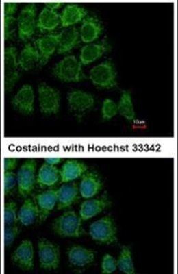
- Immunofluorescent analysis of HYOU1 in methanol-fixed Hep3B cells using a HYOU1 polyclonal antibody (Product # PA5-27655) at a 1:500 dilution.
- Submitted by
- Invitrogen Antibodies (provider)
- Main image

- Experimental details
- Immunocytochemistry-Immunofluorescence analysis of HYOU1 was performed in HeLa cells fixed in 4% paraformaldehyde at RT for 15 min. Green: HYOU1 Polyclonal Antibody (Product # PA5-27655) diluted at 1:200. Red: phalloidin, a cytoskeleton marker. Blue: Hoechst 33342 staining.

- Submitted by
- Invitrogen Antibodies (provider)
- Main image

- Experimental details
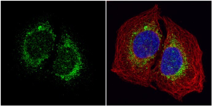
- Immunocytochemistry-Immunofluorescence analysis of HYOU1 was performed in U2OS cells fixed in 4% paraformaldehyde at RT for 15 min. Green: HYOU1 Polyclonal Antibody (Product # PA5-27655) diluted at 1:1000. Red: alpha Tubulin, a cytoskeleton marker. Blue: Hoechst 33342 staining.
Supportive validation
- Submitted by
- Invitrogen Antibodies (provider)
- Main image
- Experimental details
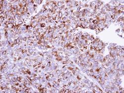
- Immunohistochemical analysis of paraffin-embedded CL1-0 xenograft, using ORP150 (Product # PA5-27655) antibody at 1:100 dilution. Antigen Retrieval: EDTA based buffer, pH 8.0, 15 min.

- Submitted by
- Invitrogen Antibodies (provider)
- Main image

- Experimental details
- HYOU1 Polyclonal Antibody detects ORP150 protein at cytoplasm on mouse fore brain by immunohistochemical analysis. Sample: Paraffin-embedded mouse fore brain. HYOU1 Polyclonal Antibody (Product # PA5-27655) diluted at 1:500. Antigen Retrieval: EDTA based buffer, pH 8.0, 15 min.
